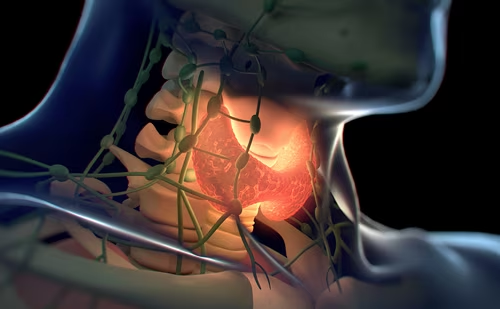
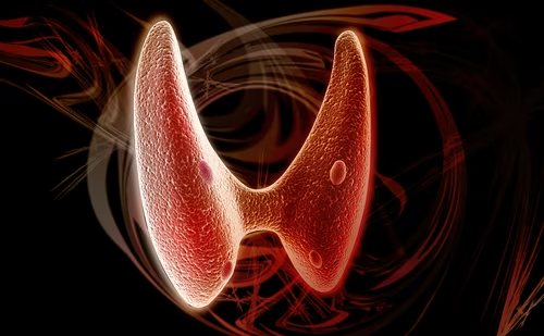
Types and Basic Biologic Features of Thyroid Cancer
Differentiated Thyroid Cancer
Types and Basic Biologic Features of Thyroid Cancer
Differentiated Thyroid Cancer
Thyroid cancer is the most common endocrine malignancy. It comprises several distinct tumor types; including papillary thyroid cancer (PTC); follicular thyroid cancer (FTC); and Hürthle cell thyroid cancer (HTC), which are tumors of the thyroid follicular cell derived from the embryonic foregut. They ordinarily concentrate iodine and sometimes synthesize and secrete thyroid hormone, and for this reason are collectively referred to as differentiated thyroid cancer (DTC). The three tumor types represent 80%, 11%, and 3% of all thyroid cancers, respectively, and have 10-year mortality rates of approximately 7%, 15%, and 25%, respectively.1
Sodium-iodide Symporter (NIS)
Many of the biologic features of normal thyroid follicular cells have been exploited to play a role in the diagnosis and treatment of DTC. The sodium-iodide symporter (NIS), which actively pumps iodine into thyroid follicular cells, has been cloned and found in DTC, as well as other benign and malignant cells.2–4 It is an integral part of the cell membrane, which, when activated by thyrotropin (TSH), initiates the first step in thyroid hormone synthesis, resulting in iodination of tyrosine molecules on thyroglobulin (Tg). This large (670kDa) glycoprotein, which is composed of two identical subunits, is released along with thyroid hormone from normal and malignant follicular cells in response to TSH.
NIS is not only present in well-differentiated thyroid cancers, but is also found in other tissues such as the lacrimal ducts, salivary glands, gastric mucosa, and the lactating mammary gland, which accounts for some of the adverse effects of radioactive iodine (131I) therapy.4–6 NIS in these non-thyroidal tissues still provides a novel opportunity to treat other malignancies. For example, most breast cancers (80%) contain NIS that can be manipulated to facilitate 131I uptake.7,8 Conversely, NIS can be selectively downregulated in breast tissue7,8 and thyroid tissue9 and can be manipulated in vitro to enhance its effect in thyroid cancer.6,10,11 These fundamental biologic mechanisms have all been exploited in the diagnosis and treatment of thyroid cancer12 and theoretically offer promise for the treatment of breast and other types of cancer.13
Medullary Thyroid Cancer
Medullary thyroid cancer (MTC) arises from thyroid C cells that are normally found in the upper third of the thyroid lobes in and around the normal thyroid follicle. The C cells, which are derived from the embryonic neural crest, secrete calcitonin in response to an increase in serum calcium or pentagastrin.
MTC comprises only 4% of all thyroid cancers. Approximately 25% of the cases are inherited as part of the multiple endocrine neoplasia type-2 (MEN-2) syndromes. The most common is MEN-2A characterized by bilateral MTCs and pheochromocytomas, causing potentially deadly attacks of hypertension and tachyarrhythmias that are often more lethal than the MTC. Another is MEN-2B associated with bilateral MTC, mucosal neuromas mainly involving the gastrointestinal (GI) tract and eyelids, bilateral pheochromocytomas, and a marfanoid habitus and is also sometimes associated with Hirschsprung disease.14,15 A third syndrome is familial MTC (FMTC), which features only bilateral MTC without other tumors. This may be confused with MEN-2A that has late onset pheochromocytoma.16
Incidence and Mortality Rates
Thyroid cancer is an uncommon disorder; however, it is an important problem, affecting approximately the same number of people as ovarian cancer or Hodgkin’s disease does yearly. In 2000 it was diagnosed in almost 123,000 people around the world, causing an estimated 8,570 deaths.17Worldwide, it afflicts approximately one in every 100,000 people each year, but its incidence is much higher in Europe and North America.17 In the US, for example, an estimated 25,690 new thyroid cancer cases will occur in 2005, accounting for almost 2% of all the new cancers in the country, and it will cause approximately 1,490 deaths.18 While it occurs at all ages, thyroid cancer is particularly common in women, occurring at approximately three-fold the rate of that in men. Its incidence peaks in mid-life in women and approximately two decades later in men.18 This is a rare but important cause of cancer in children that affects boys and girls under the age of 10 at about the same rate, but is typically discovered at a more advanced tumor stage at the time of diagnosis than that found in adults. In fact, approximately 25%19 to 50%20 of children have lung metastases at the time thyroid cancer is diagnosed compared with fewer than 10% of adults with metastases.
Historical Changes in Incidence Rates
The incidence rates of thyroid cancer in many places worldwide have increased significantly over the past three decades.17 The rates in the US have more than doubled between 1973 and 2001, varying from approximately three to seven cases per 100,000 people (P<0.05), which was due almost exclusively to a rise in the incidence of PTC without a significant change in that of FTC.21 A study from France22 identified similar changes between 1978 and 1997 during which PTC increased by 6.2% per year in men and 8.1% per year in women. Similar observations are reported from England,23 Canada,24 and other countries,25–28 but not in every part of the world.17
The increased incidence of PTC is partly due to the exposure of children and young adults to ionizing radiation,23,29–33 but has also been attributed to iodine prophylaxis,28 to alterations in histologic diagnostic criteria of clinically unimportant thyroid neoplasia,26 and to the increasingly common discovery of asymptomatic small cancers on imaging studies, so-called ‘incidentalomas’.34 A French35 study found that the increased incidence in thyroid cancer was mainly due to PTC microcarcinomas (43% of operated cancers between 1998 and 2001), which was attributed to a significant increase in the diagnostic use of thyroid ultrasonography (3% to 85%), and fine needle aspiration biopsy (8% to 36%) in the evaluation of thyroid nodules.
Historical Changes in Mortality Rates
Despite the significant rise in the incidence of thyroid cancer, its mortality rates have declined by almost 50% in the past three decades, mainly as the result of a fall in mortality rates for PTC, which has been most apparent in patients aged 40 and older at the time of diagnosis—the group most likely to die of cancer.21 Why this occurred is unknown, but is likely due to earlier treatment of less advanced tumors in recent decades. This is supported by differing incidence and mortality rates in men and women.
Mortality Rates in Men and Women
There are striking differences in the incidence and mortality rates of thyroid cancer in men and women. In women it is the most rapidly rising cause of cancer in the US, although its mortality rates have declined by over 30% in the past three decades, more than that of almost any other cancer.21 In sharp contrast, mortality rates of thyroid cancer in men during the same period have risen by nearly 4% and are two-fold those of women, largely because men present at an older age with more advanced tumors than women, making it the most rapidly rising cause of male cancer death in the US.21 This is due to late diagnosis and delayed therapy, reflecting how men utilize the healthcare system. Leenhardt et al.,34 for example, found that the proportion of women referred for evaluation of a thyroid nodule in France has increased over the past two decades, which did not happen in men—a finding which they attributed to two factors; firstly that thyroid disorders are more common in women, and secondly that women are the main consumers of healthcare.36 Men therefore die of thyroid cancer at twice the rate of women because the diagnosis and treatment are delayed and the tumor has often spread beyond the thyroid.
Genetic Alterations Leading to Thyroid Cancer
Over the past several decades, an understanding of the biology and clinical features of thyroid cancer has increased considerably. During this time clinicians have nearly perfected surgical and medical therapies for most thyroid cancer patients and have honed diagnostic paradigms that uncover residual tumor earlier now than has been possible before. Basic investigators have unlocked important secrets about the biology of these tumors, which are now being translated into new targeted treatments.
Tyrosine Kinase
The human genome contains approximately 43 tyrosine kinase (TK)-like genes and 90 protein tyrosine kinases (PTKs), which are enzymes that catalyze the transfer of phosphate from adenosine triphosphate (ATP) to tyrosine residues in polypeptides, the products of which regulate cellular proliferation, survival, differentiation, function, and motility.37 The enormous clinical importance of TKs, which are now regarded as oncogenes, suddenly became clear with the success of imatinib mesylate (Gleevec©), an inhibitor of the breakpoint cluster region-abelson (BCRABL) TK in chronic myeloid leukemia that provided the first breakthrough in targeted cancer chemotherapy.37 There is promise for similar TK-targeted chemotherapy for thyroid cancer.
Rearranged During Transfection (RET)/PTC Oncogene
Genetic alterations play an important role in the pathogenesis of thyroid cancer.A single gene located on chromosome 10, the rearranged during transfection (RET) protooncogene, codes for a cell membrane TK receptor, which is normally not expressed or is only present in very low levels in thyroid follicular cells. RET TK is activated in PTC via a chromosomal rearrangement that results in fusion of the 3’ portion of the RET gene coding for the TK domain to the 5’ portion of various genes, forming chimeric oncogenes named RET/PTC. The two most common rearrangements, RET/PTC1 and RET/PTC3,38,39 are formed by paracentric inversions of RET and its respective fusion partner, H4 or ELE1, which reside on the long arm of chromosome 10. This activates the RET receptor, which is critical to RETmediated transformation and dedifferentiation, primarily through the Ras-Raf-mitogen-activated protein kinase (MAPK) pathway.40
There is strong evidence that RET/PTC is a key first step in the formation of thyroid cancer.41–45 Transgenic mice with thyroid-targeted expression of the RET/PTC146 and RET/PTC347 develop PTC before birth. Intrachromosomal recombination events, particularly paracentric inversions, appear to be a fundamental mechanism of radiation-induced thyroid carcinogenesis. For example, RET/PTC3 rearrangements have been found in 66% to 87% of the post-Chernobyl tumors in children.48–51 Nikiforov et al.52 recently proposed that the spatial contiguity of RET and H4 may provide a structural basis for the generation of RET/PTC1 and RET/PTC3 by allowing a single radiation track to produce a double-strand break in each gene at the same site in the nucleus. RET/PTC oncogenes still do not seem to be specific to radiation-induced cancers, since they are also found in approximately 40% of sporadic pediatric PTCs and in 15% to 20% of adult cases, with considerable regional variability ranging from 5% to 40%.
B – raf Oncogene
In the past two years there has been an explosion of new information regarding mutations of B-raf, a serine-threonine kinase that maps to chromosome 7q.The three isoforms of Raf in mammalian cells are A-raf, B-raf, and C-raf that (after stimulation by Ras) activate MAPK, which is critical in the transmission of signals generated after ligand binding of membrane TK receptors.40 B-raf has the highest affinity for MAPK kinase and is the predominant isotype in thyroid follicular cells.
First reported in malignant melanomas and in a small subset of colorectal and ovarian cancers, B-raf mutations were subsequently reported in 50% to 70% of adult PTCs,53–56 all of which harbor the specific B-raf point mutation V600E (formerly designated V599E). This is now recognized as the most common genetic mutation in PTC, producing an active TK that transforms National Institutes of Health 3T3 (NIH3T3) cells with higher efficiency than the wild-type TK, consistent with its function as an oncogene.40 This B-raf mutation is also found in anaplastic thyroid cancer but not in FTC, HTC, or MTC,56 and is rarely found in children, even in those exposed to radiation.57 It is therefore considered a feature of sporadic, non-radiation-associated PTC and anaplastic thyroid cancers (ATCs).40 A few B-raf mutations have still been detected in radiation-induced thyroid cancer in children.58
Recently, a new thyroid oncogene arising via recombination of the B-raf gene was discovered in a small fraction of radiation-induced PTCs.59 The mutation involves a paracentric inversion of chromosome 7q that results in fusion between exons 1-8 of the AKAP9 gene and exons 9-18 of B raf.40 The fusion protein contains the protein TK domain and lacks the auto-inhibitory N-terminal portion of B-raf.59 It has high TK activity and transforms NIH3T3 cells, confirming its oncogenic properties.60 The adhesion- and degranulation-promoting adaptor protein nine (ADAP9) B-raf fusion was found in radiation-induced PTCs developing after a short latency, whereas B-raf point mutations were absent. The study suggests that chromosomal inversions represent a common genetic theme of radiation-associated thyroid cancer.40
Much of this previously mentioned information is likely to reach clinical practice over the next few years. For example, recent studies have shown that one subset of ATCs are derived from PTCs due to B-raf mutations and p53 mutations.61 B-raf point mutations have also been found in fine-needle aspiration (FNA) cytology specimens from PTC, making it useful for diagnosis.62 New drugs such as Geldanamycin (17-Allylamino-17- demethoxygeldanamycin (17-AAG)63 and Sorafenib (BAY 43-9006)64 that block TK activation are undergoing phase 2 studies in patients with metastatic thyroid cancer and hold new promise for patients with thyroid cancer as a direct result of new knowledge of the genetic features of the disease.
Contemporary Treatment of DTC
Thyroid Remnant Ablation
The majority of patients with DTC are treated with total or near-total thyroidectomy although small amounts of thyroid tissue nonetheless usually remain after surgery.12 As a result, 131I therapy is almost always 3 necessary to ablate residual normal thyroid tissue and microscopic cancer that remains after surgery. This reduces tumor recurrence and in some cases cancerspecific tumor rates,12 but these benefits are difficult to demonstrate in low-risk patients.65 Prospective randomized studies are still not likely to be carried out concerning the effects of remnant ablation on tumor recurrence and survival in low-risk patients,66 and most reserve 131I remnant ablation for those with tumors larger than 1cm.12,67
Thyroid remnant ablation and the treatment of DTC with 131I requires TSH-stimulated activation of NIS to pump the isotope into follicular cells, thus causing their irradiation and death. In the past, TSH stimulation was achieved by withholding thyroid hormone for three to six weeks to elevate serum TSH levels, which produces symptomatic hypothyroidism. Currently, this is best performed by injecting patients with recombinant human TSH (rhTSH—Thyrogen©), which has been available in the US and Europe for the past five years.12,68 Originally approved for diagnostic studies only, the drug has been approved in Europe and is under review for approval in the US for remnant ablation. Preparing patients for remnant ablation with this drug provides a TSH stimulus similar to that of thyroid hormone withdrawal, without producing any of the adverse effects of hypothyroidism, and is associated with less total body irradiation than that which occurs with thyroid hormone withdrawal.
Treatment of Metastases
The treatment of metastases may require surgery or 131I therapy, which is usually carried out after thyroid hormone withdrawal. However, in selected cases, rhTSH preparation may be necessary, for example when thyroid hormone withdrawal fails to evoke a sufficient TSH response, or when the patient cannot tolerate hypothyroidism or undergo withdrawal because of a concurrent medical illness. In other cases patients simply become weary of undergoing repeated thyroid hormone withdrawal. When this occurs, rhTSH is effective in preparing patients for 131I therapy. The responses have generally been approximately the same as those with thyroid hormone withdrawal, although prospective comparative studies have not been performed.69 In some cases thyroid hormone withdrawal may be necessary.70
Contemporary Follow-up Paradigms for DTC
Approximately 80% of patients respond to initial treatment of DTC resulting in low mortality rates, especially among those with PTC, the most common form of thyroid cancer. As a result, the prevalence of thyroid cancer is over 300,000 in the US and approximately 200,000 in Europe. Depending upon the completeness of initial therapy, recurrence rates have been as high as 20% to 30% in studies reporting long-term follow-up.12 Most authorities thus recommend lifelong surveillance. Any changes in follow-up management are likely to affect many people and have a major economic impact on care.
Surveillance paradigms have changed considerably in the last five years. Largely carried out in the past with diagnostic whole-body 131I scans (DxWBS) during thyroid hormone withdrawal, surveillance now mainly consists of neck ultrasonography and serum Tg measurements; however, studies from the US12,68 and Europe71 show that a TSH-stimulated Tg is required to detect persistent occult tumor.
Follow-up now comprises clinical examination at approximate six-month intervals after initial therapy, with neck ultrasonography and serum Tg measurements. When the serum Tg becomes undetectable during thyroid hormone suppression of TSH, most now advise measuring serum Tg after rhTSH stimulation (or after thyroid hormone withdrawal if necessary) and simultaneous neck ultrasonography—rather than diagnostic 131I whole-body scanning—a paradigm that is accepted by investigators in the US and Europe.68,71,72 The use of rhTSH is widely accepted by patients who want to avoid symptomatic hypothyroidism. Routine follow-up DxWBS performed with 2–5mCi of 131I or smaller amounts of 123I has been largely abandoned because the false-negative rate is in the range of 80%.68
Treating High Serum Tg Levels when the DxWBS is Negative
In 1988 Schlumberger et al.73 first identified a small but important subset of patients whose only manifestation of residual disease was an elevated serum Tg level, an observation subsequently confirmed by others.74,75When the Tg rises above a certain level in response to rhTSH or thyroid hormone withdrawal (the exact Tg value differs among laboratories and clinics), metastases may be found in patients who otherwise appear to be free of tumor. They generally have persistent locoregional tumors, which can be detected by neck ultrasonography, although approximately 30%68 have lung metastases that are not clinically apparent until therapeutic 131I has been administered and diffuse uptake is seen throughout both lungs on the post-treatment whole-body scan. Lung metastases are particularly likely to respond to 131I therapy in children and young adults, sometimes leading to a complete eradication of the tumor,19,73–75 whereas elderly patients with bulky tumor or metastases that have previously failed to pick up 131I virtually never respond to 131I therapy.76
Before empirically treating a patient with high serum Tg levels, it must be understood that Tg levels may spontaneously decline to undetectable levels over a period of several years after initial therapy.74,75,77 Conversely, an upward trend of serial serum Tg levels (i.e. a doubling of values) is much more likely to identify persistent tumor than a single Tg measurement. A single rhTSH-stimulated serum Tg>2 ng/mL has a positive predictive value (PPV) over time of approximately 80%, which may only become apparent after three to five years’ follow-up.74
Serum Anti-Tg antibodies (TgAb) in the specimen in which Tg is measured spuriously lowers the Tg result, effectively rendering it useless.78 This is mainly a problem of immunometric assays (IMA) that are used by commercial laboratories. Radioimmunoassays, which are less affected by TgAb interference,78 may provide Tg evidence of persistent disease. TgAb levels also slowly become undetectable over a two to four year period in patients who become disease-free.79
18Fluorodeoxyglucose Positron Emission Tomography ( 18FDG-PET) and Computed Tomography (CT)
One of the newest and most important innovations in the follow-up management of thyroid cancer is the use of 18FDG-PET/CT studies that now permit pinpoint detection of tumors that in the past were difficult or impossible to identify even with 18FDG-PET scans used alone. This new technology can identify surgically amenable tumors.80 Tumor definition is enhanced when the 18FDG-PET is carried out under thyroid hormone withdrawal or rhTSH-stimulation.81 18FDG tumor uptake also gives powerful prognostic information that can help guide clinical decisions.82 In the US, Medicare and other insurance carriers usually provide reimbursement for this study only when the serum Tg is elevated and a whole-body 131I scan is negative.
MTC
Astounding scientific discoveries over a period of three decades identified C-cell hyperplasia as a forerunner of familial MEN-2 syndromes and showed that calcitonin was secreted by MTC, which could be used in the diagnosis of the tumor and in evaluating the completeness of surgery, particularly when calcitonin is stimulated by pentagastrin or calcium. In the 1990s a number of activating point mutations of the RET oncogene were discovered in MEN-2 families, after which genetic testing became the basis for making therapeutic decisions in affected members of MEN-2 kindreds.
Subsequent research found a relationship between each specific RET mutation and its clinical phenotype,83 providing a basis for performing surgery in asymptomatic carriers as young as six months.16,84 However, enough discrepancies exist among the recommendations for the timing of surgery in affected children85–87 to ensure that further clinical studies are necessary to clarify this important issue for each of the specific mutations known to cause familial MTC.14
Surgery and external beam radiotherapy (EBRT) are the mainstays of treatment of MTC. The 10-year mortality rate for sporadic MTC is approximately 25%,1 which is significantly lower than that in familial cases discovered by genetic testing.87 Mortality rates are highest in sporadic MTC cases because patients usually present at an older age, with larger and more advanced tumors than those found in familial cases identified by genetic screening.
ATC
ATC is arguably the worst cancer to afflict mankind. Most cases arise in patients with long-standing benign thyroid tumors or low-grade DTC. Treatment is essentially an emergency because the tumor progresses so rapidly. Although new treatment paradigms have had some impact on survival rates, and a rare patient can be cured, many die within months of the diagnosis, and the vast majority die within a year. A combination of surgery, chemotherapy, and EBRT offer the best results, with approximately 10% surviving more than two years.88 In one such study,89 complete local response was observed in 19 patients. After a median follow-up of 45 months (range 12 to 78 months), seven patients survived and were in complete remission, of whom six had initially undergone complete tumor resection. The overall survival rate at three years was 27% and median survival was 10 months.
Conclusion
For the first time in decades, major changes in diagnostic and treatment paradigms are being seen that are being developed as the result of new technology and an enhanced understanding of the basic genetic pathways of thyroid cancer. Future developments promise exciting new advances in both the diagnosis and treatment of this diverse group of tumors.■


