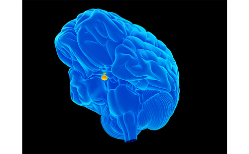
Surgery is first-line therapy for functioning pituitary adenomas (FPAs) and non-functioning pituitary macroadenomas (NFPAs) apart from prolactinomas. About 30% of NFPAs recur within five to 10 years1 and up to 60% of FPAs are still biochemically active after surgery.2–4 Somatostatin analogues and/or the growth hormone (GH)-receptor antagonist pegvisomant are often needed to achieve disease control in acromegaly. Pegvisomant is highly effective but does not control tumour growth.
Surgery is first-line therapy for functioning pituitary adenomas (FPAs) and non-functioning pituitary macroadenomas (NFPAs) apart from prolactinomas. About 30% of NFPAs recur within five to 10 years1 and up to 60% of FPAs are still biochemically active after surgery.2–4 Somatostatin analogues and/or the growth hormone (GH)-receptor antagonist pegvisomant are often needed to achieve disease control in acromegaly. Pegvisomant is highly effective but does not control tumour growth. Currently, for residual Cushing’s disease, medical treatment can only partly control excess cortisol. Dopamine agonists are first-line therapy for prolactinomas as they efficiently control both tumour size and hypersecretion. Operative treatment of large macroprolactinomas is frequently associated with complications.5,6
Pituitary adenomas that grow or are hormonally active despite surgical and medical interventions are a treatment challenge. If possible, which often is the case when located within the sella, recurrent or residual tumours are re-operated on. Management has otherwise largely depended on conventional radiotherapy (RT). With RT, long-term tumour control is excellent, while the effects on hormonal overproduction are less consistent and may take several years to become apparent.7–10
RT induces irradiation of surrounding healthy tissues, which is a safety concern. Hypopituitarism is seen in up to 50%10,11 of patients and there is increased risk of optic nerve damage, blindness, cerebrovascular disease, secondary brain tumours and dementia.12–14 It has been indicated that excess mortality in acromegaly could be related not only to poor disease control but also to RT.15,16
With the development of modern high-precision (i.e. stereotactic) techniques, irradiation of surrounding healthy tissues can be avoided.9,10 This has resulted in renewed interest in RT as a treatment option for pituitary adenomas. Localised irradiation is achieved by shaping the radiation beams to conform to the tumour shape, which spares more surrounding healthy tissues.10 Immobilisation, imaging and treatment delivery is improved compared to conventional RT.
Stereotactic irradiation can be given in one single fraction, called radiosurgery (RS), using a multiheaded cobalt unit (gamma knife RS) or a linear accelerator (Linac RS). Alternatively, it can be given in several fractions using a linear accelerator, called fractionated stereotactic radiotherapy (FSRT).10 The use of RS is restricted to smaller tumours located away from the optic chiasm and nerves, in contrast to FSRT. The use of FSRT as adjuvant therapy for different pituitary adenomas in terms of possible adverse effects and efficacy is discussed in this article.
Effects of Fractionated Stereotactic Radiotherapy
Acute and Late Adverse Effects
Only mild grade I acute adverse effects have been described with the use of FSRT. These may occur in up to 67% of patients.17 They include headache, transient local hair loss at the beam entrance site, taste/smell sensation, tiredness, eye-irritation, visual sensation, nausea and allergy to the fixation mask.17,18 No clinically apparent late adverse effects associated with conventional RT – such as neurocognitive dysfunction, cerebrovascular morbidity, mortality or secondary brain tumours – have so far been reported after FSRT.17,18 To date, there are only a few studies that have systematically investigated these factors. Longer follow-up of 10–20 years is needed to confirm that such late adverse effects can indeed be avoided with modern stereotactic techniques.
Pituitary Dysfunction
A substantial proportion of patients receiving FSRT have been operated on once or twice beforehand. For this reason, often more than 50% suffer from hypopituitarism of various degrees prior to FSRT.17,18 At a median follow-up of 60 months, new hypopituitarism was reported in approximately 20% of patients receiving FSRT.10 In heavily pre-treated subjects, 40% developed hypopituitarism at a median follow-up of 62 months.17 New pituitary dysfunction was observed eight months after FSRT at the earliest.17 These patients often present with somewhat declining hormone concentrations over time before FSRT. In clinical practice, it can therefore be recommended to screen for pituitary dysfunction and evaluate hormonal replacement therapy six to 12 months after FSRT and thereafter annually. New pituitary dysfunction may develop more than five years after FSRT.17
Vision
An obvious advantage of FSRT is that tumours close to the optic chiasm can be treated. Colin et al.19 reported that none of the patients had radiation-induced impairment of vision after FSRT. Minniti et al.18 reported that 20% of patients with field deficits improved, while three patients developed field deficits because of tumour recurrence. One patient had minimal deterioration of temporal fields without tumour growth 12 months after treatment. This remained stable during follow-up. 18 Schalin-Jäntti et al.17 reported that 36% of patients with visual field defects prior to FSRT improved, including one patient in whom the optic chiasm was not distinguishable before FSRT. The vision of none of the patients deteriorated.17 In comparison to these results, in one RS study vision did not deteriorate further, but there were no improvements afterwards.20 In another RS study, there was a 6.9% decline in visual function.21 A summary of series on FSRT is presented in Table 1.
Taken together, FSRT is not associated with radiation-induced impairment of visual acuity or vision fields. Prior vision field deficits may even improve, in contrast to RS.
Control of Tumour Growth
FSRT efficiently prevents further tumour growth of both FPAs and NFPAs (see Table 1).17–19,22–28 At a median follow-up of 39 months (range 10–60 months), control rate was 98%, which is similar to large series treated with conventional RT.10 Most series in Table 1 include patients who received FSRT as first treatment or routinely after primary surgery, which should be taken into account when comparing different studies.
In a study including only treatment-resistant NFPAs (n= 20) and FPAs (n=10), control rate was 100% at a median follow-up of 5.25 years.17 A transient, symptomless increase in tumour volume of 67%, best characterised as oedema, was observed on magnetic resonance imaging (MRI) in one patient at six months. At 19 months, the tumour volume had decreased by 29% in comparison to pre-treatment volume.17 Transient increases in tumour size six months after radiotherapy have been reported in up to 21% of patients receiving RS.29
Only a few studies have actually evaluated the magnitude of the response to FSRT (progressive disease, stable disease, partial response, complete response) by determining tumour volume or largest tumour diameter before and after treatment on serial MRIs. Colin et al.19 reported that 1.33% had progression, 9% stable disease, 89% an objective tumour response and 36% complete response at 82 months of follow-up. Schalin-Jäntti et al.17 reported that 30% had stable disease, 60% partial and 10% complete response at 62 months of follow-up. The greatest decreases in tumour volumes are observed within one to two years after FSRT.17
For clinical purposes, it can be recommended the first MRI control is scheduled one year after FSRT. The interval can thereafter be doubled if tumour control is good. Histological examination of MIB-1 proliferation index is useful for identifying potentially aggressive pituitary adenomas,30 which warrant closer follow-up as they usually grow quickly after primary surgery and may continue to do so despite FSRT. A proliferation index <5% is compatible with non-aggressive behaviour and good response to FSRT,17 while 10–30% is clearly abnormal.
Hormonal Control
Acromegaly
Milker-Zabel et al.23 reported that 16 out of 20 acromegalic patients achieved disease control after FSRT at 26 months, but it is unclear how this was defined because GH concentrations after FSRT were not reported. According to Giustina et al.,31 biochemical control is defined as mean GH <2.5ug/l, nadir GH in reponse to an oral glucose tolerance test <1.0ug/l and normal age- and sex-adjusted insulin-like growth factor 1 (IGF-1). Minniti et al.18 reported a 45% decrease in basal GH levels. Disease control was achieved in six out of 18 patients (35%) who had stopped taking somatostatin analogues at a median follow-up of 39 months. Roug et al.28 reported that 50% of 34 patients were biochemically controlled 30 months after FSRT, 10 of whom were off somatostatin analogues.
It is important to achieve disease control in acromegaly, as this decreases morbidity and mortality. With somatostatin analogues, it is possible to control tumour growth and GH and IGF-I levels decrease in approximately 90% of patients.32–34 Despite this, GH levels <2.5μg/l are only achieved in about 50% of cases.32–34 In persistently active cases disease control is probably best achieved by efficiently combining different treatment regimens, including somatostatin analogues, dopamine agonists and/or pegvisomant.
FSRT can serve as valuable adjuvant therapy in treatment-resistant cases, especially in cases with large residual tumours located outside the sella, which are therefore not suitable for further surgery. FSRT can shrink the residual tumour and decrease excess hormonal secretion.
In addition to improving disease control in treatment-resistant cases, FSRT may also decrease the need for expensive somatostatin analogue17 and pegvisomant treatment. Pegvisomant, the most efficient medical treatment for acromegaly, does not control tumour growth and only IGF-1 can be used for assessment of disease control. It is also the most expensive treatment and, as with somatostatin analogues, must be administered parenterally.
Cushing’s Disease, Large Prolactinomas and Follicle-stimulating Hormone Adenomas
There are only limited data on FSRT in corticotroph adenomas, large prolactinomas and gonadotropinomas.
In a series of 12 patients with Cushing’s disease, control of elevated cortisol was reported in 75% at a median follow-up of 29 months.19 Treatment options other than surgery for severe Cushing’s disease include medical therapy (which partly controls cortisol excess), bilateral adrenalectomy and, in selected cases, temozolamide chemotherapy.35,36
For large prolactinomas, it is essential to achieve both tumour and hormonal control. Dopamine agonists are highly efficient but may in rare instances not be sufficient alone. They may also be associated with adverse events or the patient may be non-compliant. In three patients with giant (>4cm) progressive prolactinomas on dopamine agonists, a remarkable decrease in tumour size of 65–100% and hormonal control was observed after FSRT at 30–105 months of followup. 17 In a patient with a growing macroprolactinoma previously operated on and not compliant with cabergoline treatment, FSRT resulted in complete tumour response and a 94% decrease in prolactin at 80 months.18
Larger studies are clearly warranted to confirm these findings. In one patient with an follicle-stimulating hormone (FSH)-adenoma, further tumour growth was prevented. FSH-secretion decreased by more than 75%.17
Conclusions
The primary indication for FSRT is NFPAs and FPAs resistant to surgery and/or medical treatments. FSRT is safe and efficient, especially in controlling tumour growth, but also has well-documented effects in acromegaly. Data on Cushing’s disease are scarce. Results from a small series of large prolactinomas are promising. FSRT is also suitable for larger pituitary tumours close to the optic chiasm, in contrast to RS, and seems to be at least as efficient. There are, however, fewer published data and shorter follow-up of patients who have received FSRT compared with RS and there are no direct comparisons.
The technique chosen by clinicians probably mostly depends on availability, but conventional RT should now be avoided, bearing in mind the long-term complications. Longer follow-up of 10–20 years will confirm whether the late adverse effects associated with conventional RT are avoided with modern high-precision stereotactic techniques.


