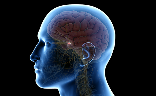
The ability of an organism to respond to stressful stimuli is fundamentally important to that organism’s continuing survival. Recognition of a stressor elicits a range of physiological changes that enable the organism to cope and to facilitate the restoration of homeostasis. Many of these physiological changes are mediated via activation of the hypothalamo–pituitary–adrenocortical (HPA) axis and the consequent secretion of glucocorticoids (GCs) by the adrenal gland.
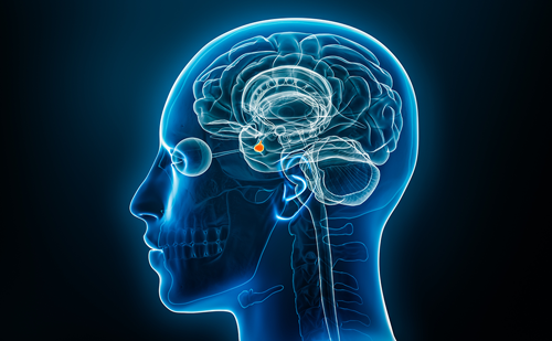
The ability of an organism to respond to stressful stimuli is fundamentally important to that organism’s continuing survival. Recognition of a stressor elicits a range of physiological changes that enable the organism to cope and to facilitate the restoration of homeostasis. Many of these physiological changes are mediated via activation of the hypothalamo–pituitary–adrenocortical (HPA) axis and the consequent secretion of glucocorticoids (GCs) by the adrenal gland. Stimulation of the HPA axis is triggered by neural and humoral mechanisms that converge on the parvocellular neurones in the hypothalamic paraventricular nucleus (PVN) and cause release of corticotrophin-releasing hormone (CRH) and arginine vasopressin (AVP) into the hypothalamo–hypophyseal portal complex for transportation to the anterior pituitary gland. Here, these neurohormones bind to specific CRH and AVP receptors (CRH-R1 and V1b, respectively) on corticotroph cells to induce the release of corticotrophin (adrenocorticotrophic hormone [ACTH]) into the systemic circulation. ACTH acts within the adrenal glands to increase the synthesis and release of GCs, cortisol (in man and other primates) and corticosterone (in rodents). The secretion of these steroid hormones is further regulated by complex negative feedback effects of the GCs themselves on the pituitary gland, hypothalamus and extra-hypothalamic centres in the brain (e.g. hippocampus, brainstem). GCs were originally named on the basis of their influence on metabolic processes, specifically the generation of glucose from protein and lipids. However, GCs also exert a plethora of effects that together serve to maintain homeostasis. GCs thus prepare the organism to respond to stress and also protect the organism from the stress itself, in part by limiting the pathophysiological responses (e.g. inflammation) to the stress that, if left unchecked, may themselves threaten homeostasis.1 Glucocorticoids Regulation of Plasma and Intracellular Glucocorticoid Levels
How do stressful stimuli activate the HPA axis and thus precipitate GC secretion? The PVN is the key site within the brain where many stress-sensitive ascending and descending neural pathways converge and trigger HPA activation. Fibres originating in the brainstem, prefrontal cortex, hippocampus, raphe nucleus and amygdala are particularly important in this regard. These pathways use a range of neurotransmitter/neuromodulator substances to modulate the secretion of CRH and AVP, including acetylcholine, noradrenaline, 5-hydroxytryptamine, gamma-aminobutyric acid, neuropeptide Y, endogenous opioids and various growth factors and cytokines. However, while categorically distinct stressors (i.e. physiological versus emotional) use distinct and specific pathways and transmitters, they recruit largely the same select group of genes within the PVN2 to induce release of CRH and AVP and, thus, secretion of GCs. Secretion of GCs into the circulation occurs in a pulsatile and circadian fashion. Pulse frequency is approximately one to three pulses per hour3 in man; variations in pulse amplitude over the 24-hour cycle underpin the circadian profile of maximal GC levels in the morning, prior to awakening (approximately 800nM), and low levels in the evening (approximately 200nM).4–6 Plasma cortisol levels are further increased by stress and, depending on the nature and intensity of the stress, may rise as much as ten-fold above basal levels.1
GCs in the bloodstream are largely bound to plasma proteins (~90%), in particular cortisol-binding globulin (CBG).7Only the free steroid can cross cell membranes and gain access to the intracellular receptors that mediate the biological effects of the steroids. Therefore, binding to plasma proteins limits the access of circulating GCs to their receptors by restricting entry to target tissues. However, in certain conditions (e.g. inflammation), cortisol may be released from CBG in the target tissues by the actions of human leukocyte elastase, which cleaves CBG.8 In addition, some tissues possess membrane-bound CBG receptors, which can internalise both the binding protein and the associated cortisol (see Figure 1).9
Two further mechanisms determine the bioavailability of free cortisol within the cell. The first, termed pre-receptor ligand metabolism, is mediated by two intracellular enzymes, 11β- hydroxysteroid dehydrogenase 1 and 2 (11β-HSD1 and 11β-HSD2), which regulate the interconversion of cortisol and its biologically inert metabolite, cortisone. 11β-HSD1 acts as a reductase and thus regenerates bioactive cortisol from inactive cortisone and increases the local cortisol concentration. Conversely, 11β-HSD2 catalyses the conversion of cortisol to cortisone and thus reduces the availability of cortisol within the cells. These two enzymes are expressed in a highly tissue-specific manner. 11β−HSD1 is particularly prevalent in GC-responsive metabolic tissues such as the liver and central nervous system,10,11 while 11β-HSD2 is predominantly located within the kidney and protects high-affinity mineralocorticoid receptors from cortisol.10,12 The second mechanism is the multidrug-resistant drug (mdr) transporter protein, P-glycoprotein, which is also expressed in a highly tissue-specific manner and exports cortisol from cells, thus reducing the intracellular concentration of the steroid. The tissue-specific patterns of expression of 11β-HSD1, 11β-HSD2 and P-glycoprotein thus provide effective mechanisms for local regulation of the access of GCs to their receptors.
Mechanism of Glucocorticoid Action
GCs act mainly via intracellular receptors, of which there are two main types: the mineralocorticoid receptor (MR) and the GC receptor (GR). These receptors mostly act as transcription factors, regulating the expression of specific target genes. The number of target genes is large – possibly as high as 1% of the genome. MR is a high-affinity receptor that cannot distinguish cortisol from the mineralocorticoid aldosterone. The MRs have a highly tissue-specific pattern of expression, and in tissues classically associated with aldosterone actions (e.g. kidney) are protected from cortisol by 11β−HSD2. By contrast, the GR is a low-affinity receptor with a high specificity for GCs. It is widely distributed in the body. In many tissues, particularly those associated with metabolism (e.g. liver), access of cortisol is facilitated by 11β-HSD1. GRs are cytoplasmic receptors that, in the absence of ligand, exist in a complex with accessory proteins, such as heat shock proteins, which act as chaperones to retain the GR within the cytoplasm.13 Binding of cortisol to the ligand-binding domain within the C-terminal of GR14 induces a conformational change that promotes the dissociation of the heat shock proteins, exposure of the nuclear localisation signal and translocation of the ligand–receptor complex to the nucleus via an importin-mediated mechanism.15 Ligand-bound GR uses two principal mechanisms to influence transcription of specific target genes: transactivation and transrepression. Transactivation requires homodimerisation of GR subunits and interaction of the GR DNA-binding domain with conserved GC response elements within the promoter region of responsive genes,16 a process facilitated by the recruitment of transcriptionally active proteins.14 Interestingly, it appears that small changes in the DNA recognition sites for GR can subtly alter GR transcriptional activity, suggesting that there may be gene-specific GR effects within tissues.17 GR-induced transrepression occurs principally via a mechanism independent of DNA binding,18 with GR monomers specifically binding to and interfering with the actions of transcription factors such as nuclear factor kappa B (NF-κB) or activating protein- 1 (AP-1).19 For example, the ability of the NF-κB p65 subunit to induce expression of pro-inflammatory mediators is suppressed by binding of GR.20 These differences are highly cell-specific and can determine GC responses, with specific genes demonstrating activation or repression depending on circumstances.
In addition to influencing gene transcription directly, GCs may also act via non-genomic mechanisms.21,22 For example, GCs promote the cellular exportation of the anti-inflammatory protein annexin 1 from pituitary folliculostellate cells,23,24 predominantly through a non-genomic mechanism.24 Croxtall and colleagues demonstrated that this action involves the rapid release of Src kinase from cytoplasmic GR heterocomplexes and subsequent inhibition of arachidonic acid release.25 It is also possible that some non-genomic actions are associated with activation of a membrane-bound GR. These receptors are present in small numbers per cell, but are actively upregulated after immunostimulation. It has been suggested that overstimulation of the immune system would lead to upregulation of membrane-bound GR, which would act in a feedback manner to reduce the excessive immune reaction.26 The nongenomic mechanisms of GC action remain poorly understood; therefore, further studies are warranted, particularly since manipulation of these events may prove therapeutically useful.
Glucocorticoids and Human Disease
The clinical features associated with conditions of severe GC excess (Cushing’s syndrome) and deficiency (Addison’s disease) are well established, but these conditions are relatively rare. However, considerable evidence points to a role for GCs in the pathophysiology of numerous other endocrine-related disorders such as type 2 diabetes, dyslipidaemia and metabolic bone disease. Prolonged increases in physiological GC production are most likely to be the result of exposure to chronic stress. Alternatively, alterations in the local intracellular mechanisms that regulate the access of GCs to their receptors may cause local disturbances in GC homeostasis that influence disease processes.
Acute stress is an allostatic process that aims to restore homeostasis via adaptation, using mediators from numerous systems including the HPA axis. Chronic stress is likely to be associated with allostatic overload, where adaptive processes are used in a sustained manner. It is this prolonged inappropriate use of adaptive physiological processes that can result in dysfunction or disease. For example, increased food intake and fat deposition can be seen as an allostatic response to ensure there is sufficient metabolic resource to maintain homestatic processes, whereas in allostatic overload, this might result in abdominal obesity. Prolonged increases in cortisol due to exposure to chronic stress are likely to exact an allostatic load, with increased wear and tear apparent in certain GC-senstitive tissues, whereas decreased function will be apparent in other tissues owing to prolonged inhibitory effects of GCs or redistribution of metabolic resource to physiological systems involved in restoring homeostasis. However, it should be noted that tissue-specific alterations in GC concentrations without corresponding increases in circulating GC levels can also influence disease processes. It is interesting to note that the high circulating levels of GCs caused by Cushing’s syndrome are associated with a number of negative metabolic outcomes,10,27 whereas near normal serum GC levels are usually found in patients with the more prevalent metabolic syndrome.28 It has been suggested that an alteration in tissue sensitivity to GCs underlines the metabolic syndrome, specifically an alteration in the expression of 11β-HSD1. Numerous animal studies have demonstrated that 11β-HSD1 expression within metabolic tissues (e.g. adipose tissue, liver) is correlated with an adverse metabolic outcome,29–31 and metabolic disease within humans is commonly associated with elevated 11β- HSD1 expression/activity.32,33 Therefore, tissue-specific alterations in 11β-HSD1 expression coupled with increased intracellular GC concentrations and subsequent GR activation may be a common feature of metabolic disease. It has also been demonstrated that there are a number of polymorphisms within the GC receptor gene itself that influence GC sensitivity.34–36 Several of these GC receptor variants are associated with hypersensitivity to GCs37–39 and therefore might predispose an individual to negative health outcomes associated with GC overexposure.The developing organism is particularly sensitive to GCs, and unwanted increases in foetal GR activation due to maternal stress or synthetic GC administration (often used in peri-natal medicine to mature the lung in conditions of pre-term birth) have the potential to induce programming effects on multiple body systems. Studies performed on laboratory animals have shown that exposure of the developing foetus or neonate to supraphysiological GC levels or synthetic GCs results in irreversible morphological and physiological changes in the organism, which predispose it in adulthood to diseases that are endemic in the developed world, such as type 2 diabetes, cardiovascular disease, depression and other mental health disorders. More limited data from clinical studies support these conclusions. The maternal–foetal unit is designed to prevent excessive foetal exposure to GCs, with placental 11β-HSD-2 acting as a barrier to the passage of maternal GCs.40–42 However, this mechanism may become saturated if endogenous GC levels rise excessively, or can be ineffective, as is the case with synthetic GCs. A key feature of GC programming in early life is prolonged, tissuespecific change in the expression of GR. Reduced GR expression within the HPA axis leads to impairment of GC negative feedback in adulthood, leading to raised GC levels and exaggerated HPA responses to stress. Conversely, GR expression in the liver is increased, thus predisposing the individual to hyperglycaemia.
Glucocorticoids and Endocrine-related Disorders
This section deals with a number of endocrine-related disorders that are associated with aberrant GC levels and in terms of pathophysiology may be linked with chronic tissue-specific alterations in GC actions.
Glucocorticoids and Hyperglycaemia/Type 2 Diabetes
Patients with Cushing’s syndrome or on long-term GC therapy classically present with hyperglycaemia43,44 and symptoms of type 2 diabetes43 – in this case termed steroid diabetes. It is important to note that the development of type 2 diabetes is usually multifactorial, but this article will discuss steroid diabtetes induced by increased GC levels. GCs act within the liver to upregulate the rate-limiting enzyme for gluconeogensis, phosphoenol-pyruvate carboxykinase (PEPCK), providing a mechanism to explain GC-induced hyperglycaemia. Insulin resistance is the major biological risk factor for type 2 diabetes,45 and is associated with both a reduced secretion of insulin by the endocrine pancreas and a reduction in insulin sensitivity within peripheral tissues. GCs decrease insulin secretion46 and also act on multiple targets to influence insulin sensitivity, downregulating components of insulin signalling such as insulin receptor substrate proteins 1 and 2,47 phosphoinositide 3 kinase activity48 and Akt phosphorylation.49 In addition, there is a strong positive correlation between GR expression levels and the degree of insulin resistance.50
The link between hyperglycaemia/diabetes and GCs is further strengthened by studies focused on GC pre-receptor metabolism. 11β-HSD-1 overexpression in mice, which increases local GC concentrations in specific target tissues, is associated with modest insulin resistance,51 whereas 11β-HSD-1 knockout mice show a reduced ability to regenerate intracellular GCs, improved insulin sensitivity,52,53 impaired induction of PEPCK, a reduced hyperglycaemic response to stress29 and an improvement in several aspects of GCinduced diabetes.54 In humans, increased HPA activity is associated with type 2 diabetes,55–57 with high circulating cortisol levels positively correlated with more severe complications from type 2 diabetes.58
Glucocorticoids and Dyslipidaemia/Obesity
GCs are important physiological regulators of energy balance. It is therefore not surprising that the development of metabolic pathologies such as obesity has been strongly associated with dysfunctional GR signalling. Cushing’s patients present with centripetal obesity, which is directly linked to excessive GC action.59 Adipocytes in the abdominal fat pads are more GR-rich than peripheral adipocytes and thus more sensitive to GCs.60 Within central fat, GCs increase pre-adipocyte differentiation and promote the prolipogenic pathways, thereby increasing cellular hypertrophy.61,62 In non-cushingoid patients the development of obesity is not necessarily associated with increased circulating levels of GCs;10 however, in some cases at least, there is evidence of altered tissue sensitivity to GCs owing to tissue-specific upregulation of 11β-HSD-1 activity.63,64 There is evidence that 11β-HSD-1 activity is impaired in the liver but enhanced in adipose tissue in obese human patients.33,65 Furthermore, the adverse metabolic complications of obesity in mice are prevented by 11β-HSD-1 gene deletion,66 whereas overexpression of the enzyme in adipose tissue results in metabolic abnormalities.67 Studies on obese and lean human individuals have demonstrated increased adipose tissue 11β-HSD-1 expression in obese subjects68 and a direct association between 11β-HSD-1 levels and metabolic abnormalities in obese women.69 However, tissue-specific overexpression of 11β-HSD- 1 in the liver is associated with numerous metabolic alterations but not with changes in fat depot mass.51 Furthermore, other authors have failed to find an association between obesity and 11β-HSD-1 activity in adipose tissue.70 Thus, while there is evidence to link tissue-specific alterations in GC bioavailability with the metabolic abnormalities associated with the development of obesity, further studies are necessary to understand fully the role of GCs.
Obesity is associated with an increased risk of coronary heart disease due partly to impaired trapping and breakdown of fatty acids by adipocytes, which facilitates atherogenic dyslipidaemia and is associated with low levels of high-density lipoprotein cholesterol, (HDL-C), elevated triglycerides and increased low-density lipoproteins (LDL). Synthetic GR agonists increase serum triglyceride levels and cause accumulation of hepatic lipid droplets in vivo, whereas disruption of GR action specifically decreases serum triglyceride in a mouse model of fatty liver.71 These GC-mediated effects are most likely due to a decrease in β-oxidation of fatty acids and increased hepatic uptake and storage of fatty acids as triglyceride. Recent studies indicate that GR activation influences the expression of multiple genes directly involved in fatty acid and triglyceride metabolism that may contribute to systemic dyslipidaemia. Enhanced GR activity is also associated with decreased pancreatic lipase activity and fatty acid β-oxidation, profound inhibition of adipose tissue lipoprotein lipase (which would normally act to increase uptake of triglyceride-derived fatty acids) and increased liver cholesterol.71–73
Glucocorticoids and Depression
Depression is a complex illness characterised by a spectrum of clinical symptoms including low mood, alterations in appetite and weight, psychomotor agitation or retardation, sleep disruption and suicidal ideation. The development of the disease is influenced by genetic and psychosocial factors as well as biological disturbances.74,75 There is undoubtedly a very strong correlation between disturbances in HPA function levels and the development of depression. Over 50% of Cushing’s patients present with depressive symptoms,76 and a similar percentage of depressed patients present with hypercortisolaemia.77 Onset of depression is correlated with stressful life events associated with prolonged elevations in circulating GCs, such as divorce or unemployment.78
Patients with major depression have been shown to exhibit increased concentrations of cortisol in the plasma, urine and cerebrospinal fluid,79,80 exaggerated cortisol responses to exogenous ACTH81 and an enlargement of both the pituitary and the adrenal glands.82 In addition, a multitude of studies have demonstrated that GC-mediated feedback inhibition of the HPA axis is impaired in depression; thus, unlike normal patients, approximately 50% of depressed patients fail to respond to synthetic GCs with a reduction in serum cortisol.83 In addition, many effective antidepressant treatments have been shown to modulate cortisol secretion.84 Indeed, the GR is now an important target for novel antidepressants, and some compounds that specifically reduce the effects of cortisol have produced successful results in clinical trials.85 Depression is associated with structural abnormalities in a number of corticolimbic structures that play important roles in cognition and emotional processing, such as the hippocampus, amygdala and prefrontal cortex.86,87 Each of the aforementioned brain regions is rich in GR,88–90 and increases in salivary cortisol induced by acute stress are associated with decreased activity within the hippocampus and amygdala.91 Interestingly, volumetric reductions in the hippocampus are observed in Cushing’s patients, and these reductions are partially reversed if the hypercortisolaemia is corrected.91–94 While these clinical data are suggestive of a link between hypercortisolaemia and depression, they do not demonstrate true causation. Animal models have attempted to demonstrate a direct link between high circulating GC levels and depressive-like symptoms. However, there is some debate as to whether animals can be classified as depressed and whether the tests used to assess the disease in animals can truly be correlated with clinical symptoms of depression, which are subjective and highly variable. Generally, a good animal model of depression will demonstrate some of the behavioural and neurochemical changes associated with the disease as well as responding to well-established antidepressant treatments. In addition, the ability to examine the aetiology of depressive illness is another favourable asset for any animal model.95
Glucocorticoids and Osteoporosis
GC treatment is associated with rapid bone loss, and fractures are a common side effect of long-term GC therapy.96 Bone remodelling is dependent on the absorption of old bone matrix by osteoclasts, followed by the generation of new bone matrix by osteoblasts that subsequently enter the resorptive lacuna. GCs interfere with bone matrix formation via induction of osteoblast apoptosis via activation of caspase-3, in addition to decreasing the number of osteoblast precursor cells available for differentiation.97 Chronic GC treatment induces the expression of macrophage colony-stimulating factor (MCSF) and receptor activator of NF-κB ligand (RANKL), both of which are necessary for osteoclast development. GCs also increase osteoclast maturation and survival, which is classically associated with increased bone resorption and rapid loss of trabecular bone.98,99
However, recent studies suggest that osteoclasts induce a more complex effect on bone remodelling. Osteoclasts act to resorb old bone, an action that requires cytoskeletal organisation. As osteoclasts absorb the old bone matrix, they release factors that promote the movement of osteoblasts into the resorptive lacuna, which then act to synthesise new bone. Long-term GC therapy suppresses specific osteoclast functions related to cytoskeletal organisation and recruitment of osteoblasts, and therefore greatly reduces new bone formation.100
Future Perspectives
GCs are predominantly used as anti-inflammatory and immunosuppressive agents. Despite the undoubted clinical benefit obtained from the use of these drugs, the side-effect profile associated with their use remains a huge problem.43 Anti-inflammatory and immunosuppressive actions of these drugs predominantly feature GC-mediated transrepression, whereas the side effects often involve transactivation. Selective GR agonists with a pharmacological action mostly based on transrepression with little effect on activation could retain the desirable clinical effects of these drugs while considerably reducing side effects.43 Animal models demonstrating tissue-specific knockout or overexpression of GC targets will also help provide a more accurate picture of GC action in vivo.
Antistress gene therapy is also a potential tool to protect against tissue impairment due to prolonged elevations in circulating GCs. Dumas and colleagues101 have recently demonstrated that tissue-specific expression of 11-β-HSD-2 within the hippocampus offsets neurophysiological disruptions induced by chronically increased GC levels. Thus, a better understanding of tissue-specific GC physiology will allow us to develop more sensitive GC-based therapies and generate treatments aimed at improving outcomes in diseases/disorders associated with chronically increased GC levels.