touchENDOCRINOLOGY touchENDOCRINOLOGY
Thyroid Disorders
Read Time: 6 mins

Coexistence of Thyrolipomatosis and Tongue Squamous Cell Carcinoma: A Case Report

Copy Link
Published Online: Jan 30th 2023 touchREVIEWS in Endocrinology. 2023;19(1):103-7 DOI: https://doi.org/10.17925/EE.2023.19.1.103
Authors: Jose Paz-Ibarra, Marcio Concepción-Zavaleta, Daniel Mendoza-Quispe, Jacsel Suárez-Rojas, Katia Rivera Fabián, Diana Deutz-Gómez, Juan Quiroz-Aldave, José Somocurcio Peralta, Tula Ayquipa Arróspide
Quick Links:
Abstract
Article
Article Information
Abstract:
Overview

Thyrolipomatosis, a diffuse non-neoplastic infiltration of fatty tissue in the thyroid gland, is an extremely rare condition with only about 30 cases reported worldwide. A few of these cases report the concurrency of thyrolipomatosis and malignant neoplasms in the thyroid or colon, but never with tongue cancer. A 44-year-old female patient with an infiltrative tongue mass suggestive of carcinoma presented for an outpatient consultation. Cervical imaging revealed multiple lymphadenopathies and a multinodular goitre with diffuse fatty infiltration, suggestive of thyrolipomatosis. Surgical intervention included partial resection of the tongue and thyroid (left hemiglossectomy and right hemithyroidectomy, respectively) and lymphadenectomy. The thyroid specimen showed diffuse fat metaplasia of the stromal thyroid tissue, confirming incidental thyrolipomatosis. During post-operative follow-up, the patient presented with recurrence of squamous cell carcinoma as indicated by new right-sided thyroid nodules, left-sided lymphadenopathies with confirmatory biopsy, and a growing neck mass that became infected. The patient developed septic shock and later died. Thyrolipomatosis causes thyroid swelling and can be clinically detected as goitres or as an incidental finding. Diagnosis is suggested by cervical imaging (ultrasonography, computed tomography or magnetic resonance), but confirmation is histological after thyroidectomy. Although thyrolipomatosis is benign, it could develop concurrently with neoplastic diseases, especially on embryologically related tissues (e.g. thyroid and tongue). This case report is the first in the literature describing the coexistence between thyrolipomatosis and tongue cancer in an adult Peruvian patient.

Keywords

Lipomatosis, tongue neoplasms, squamous cell carcinoma, goitre, Peru

Article:

Thyrolipomatosis is a rare condition defined as a diffuse non-neoplastic infiltration of fatty tissue in the thyroid gland.1 Although fatty infiltration is common in other glands (e.g. salivary glands, parathyroids, thymus and pancreas), it is rare in the thyroid gland.2 If present, it is most frequently nodular (i.e. thyrolipoma) rather than diffuse (i.e. thyrolipomatosis).3 Thus, since the first case of thyrolipomatosis was reported in 1942 by Dhayagude,4 only about 30 cases have been published worldwide.5–7 In addition, few of these cases showed concurrency of thyrolipomatosis and malignant neoplasms in the thyroid or colon, but never with tongue cancer. In this report, we present the case of an adult Peruvian patient with squamous cell carcinoma of the tongue and incidental thyrolipomatosis, along with a literature review.

Case report

A 44-year-old female patient was assessed at the head and neck surgery outpatient department at a tertiary care public hospital in Lima, Peru, in November 2021. Her main complaint was a tongue mass that had progressively enlarged over the past 1.5 years, which was associated with dysphagia to solids and weight loss. Her history included non-toxic multinodular goitre and juvenile rheumatoid arthritis that were diagnosed when she was in her twenties and treated with leflunomide. There was no history of radiation exposure or any contributing family history. Physical examination revealed a left-sided pearly mass on the tongue (3 cm × 1.5 cm) suggestive of carcinoma, goitre grade IB (i.e. palpable goitre with a painless, mobile, right-sided thyroid nodule), palpable bilateral cervical lymphadenopathies, cervical skin without changes, undernutrition (body mass index 17.8 kg/m2) and vital signs in normal range. Laboratory examinations revealed mild microcytic anaemia (haemoglobin: 11.4 mg/dL; mean corpuscular volume: 73 fL). Cervical ultrasonography revealed a predominantly left-sided multinodular goitre, a left-sided thyroid nodule (31 mm × 22 mm; mixed solid and cystic content; slightly hypoechogenic) and multiple bilateral lymphadenopathies (i.e. a right-side node of 24 mm × 5.9 mm, another right-side node of 12 mm × 4 mm, and a left-sided node of 27 mm × 11 mm). Fine-needle aspiration (FNA) of the lymph nodes revealed squamous cell carcinoma in the left adenopathy and reactive lymphoid hyperplasia in the right.

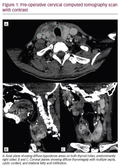
The cervical computed tomography (CT) scan with oral contrast staged the squamous cell carcinoma of the tongue as cT3 N2b Mx G1 according to TNM classification (Figure 1). The CT scan also revealed infiltration of the parotids, lymph node conglomerate in the left cervical chain with an infiltrative appearance, and diffuse thyromegaly with multiple septa, cystic content and bilateral fatty infiltration.

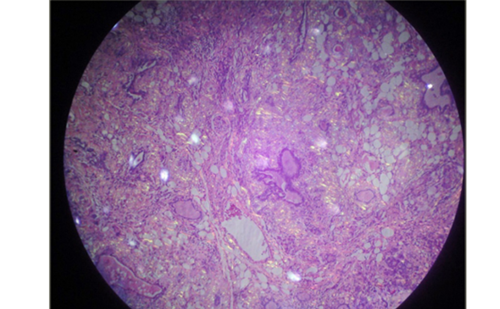
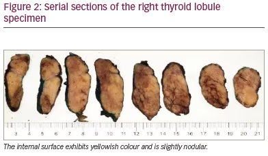
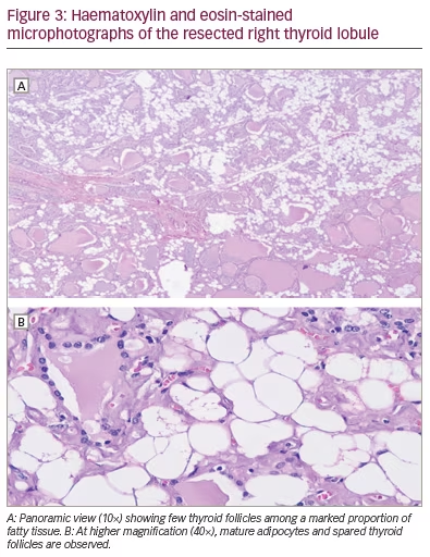
The patient was admitted for surgical intervention that consisted of partial resection of the tongue and thyroid (i.e. left hemiglossectomy and right hemithyroidectomy, respectively) alongside a radical modified cervical lymphadenectomy (type I on the left, type III on the right), and a procedure for mandibular correction (i.e. symphyseal osteotomy). Gross examination of the thyroid specimen (Figure 2) showed a reddish, right thyroid lobule, normal weighted (15 g), increased in size (6 cm × 3.5 cm × 2 cm) of elastic consistency containing colloid cysts and remaining homogeneous parenchyma. Histologic examination of the thyroid specimen (Figure 3) showed non-neoplastic macrofollicular colloidal hyperplasia with cystic degeneration and diffuse fat metaplasia of stromal thyroid tissue, confirmatory of thyrolipomatosis. Biopsy was Congo red negative, excluding amyloid infiltration.

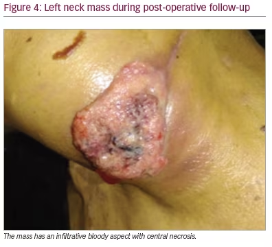
During post-operative follow-up, the patient was euthyroid (thyroid-stimulating hormone 0.733 mU/L). Three months post-operatively, a control cervical ultrasonography suggested metastasis, after images showed a left-sided thyroid nodule with an American College of Radiology Thyroid Imaging Reporting and Data System score of 4 (18 mm × 13 mm), a left-sided lymphadenopathy (10 mm × 5 mm) and a left cervical mass (43 mm × 26 mm); all of these pathologies had malignant features (e.g. ill-defined borders, heterogenous content and marked Doppler vascularity). Recurrent cancer was confirmed by a FNA cytology of the lymphadenopathy that showed squamous malignant cells. The left neck mass grew rapidly and became painful and bloody (Figure 4). However, as the referred mass was highly vascularized and was located around major vessels (e.g. left carotid artery, subclavian artery and jugular vein, according to angiotomography), the patient was a poor surgical candidate due to high risk of massive bleeding. The multidisciplinary medical team suggested palliative locoregional radiotherapy to reduce mass size and gastrostomy due to dysphagia. Five months post-operatively, the left neck mass became infected; the patient was admitted to hospital and received broad-spectrum antibiotics (meropenem and vancomycin), but septic shock progressed and the patient died.

Discussion

This case report is the first in literature documenting concurrent thyrolipomatosis and squamous cell carcinoma of the tongue in an adult Peruvian patient. This presentation suggests that, although thyrolipomatosis is a benign and incidental condition, it may be concurrent with malignant neoplasms.

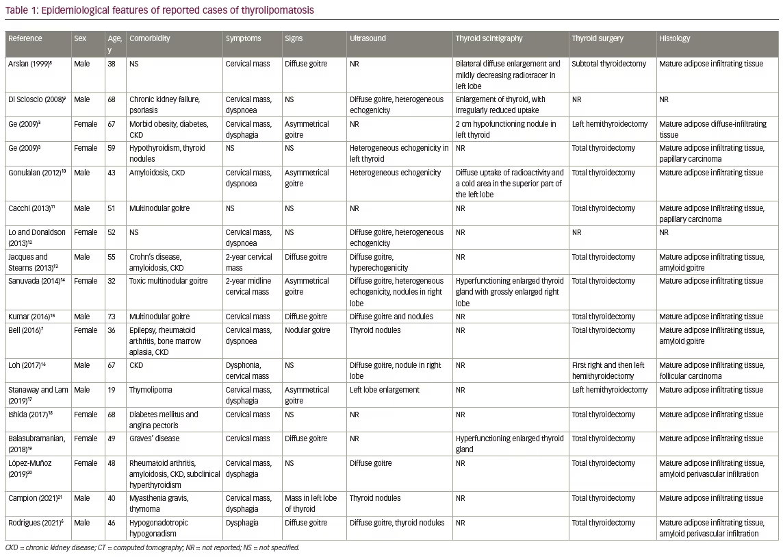
Table 1 summarizes reported cases and lists the epidemiological features of thyrolipomatosis. For example, there is no predominance by sex (56% male), its occurrence is mostly in middle-aged adults (mean age 51 years; range 44–57 years) and it is usually associated with systemic conditions like chronic kidney failure (39%) and amyloidosis (17%).3,6–21

Thyrolipomatosis is a benign condition because the fatty infiltration of the thyroid gland is diffuse but non-neoplastic. However, some case reports suggest the coexistence of thyrolipomatosis with malignant neoplasms such as papillary thyroid carcinoma or colon cancer.6,22,23  As mentioned, this report is the first published case of a patient with thyrolipomatosis with concurrent squamous cell carcinoma of the tongue. Table 1 confirms that it may coexist with malignancies in less than 20% of cases.

A hypothesis of causality between thyrolipomatosis and tongue cancer is not reasonable, since these entities have different histopathological findings; nonetheless, the organs in which both conditions are located (i.e. the thyroid and tongue) are embryologically related and may partly explain their coexistence. During the pre-natal period, the thyroid gland arises near the base of the tongue and migrates caudally; it remains attached to the tongue via the thyroglossal duct, which almost always obliterates entirely.24

The underlying pathophysiology of thyrolipomatosis is unclear; it may involve either embryological growth of fatty tissue in the thyroid gland,25 fatty metaplasia of stromal fibroblasts in response to hypoxia14 or a mutation of a mitochondrial protein.25 As thyrolipomatosis causes thyroid swelling, patients usually present with a progressive goitre, either diffuse or nodular, which may be asymptomatic or cause compressive symptoms such as dyspnoea, dysphagia or dysphonia.2,26 Our patient presented with dysphagia due to another condition (a lymphadenopathy). Most patients, as with our case, have normal thyroid function (see Table 1), although some may present with elevated14,27 or decreased 3 thyroid hormone levels.

On work-up, cervical imaging (ultrasonography, CT or magnetic resonance) can suggest adipose thyroid tissue infiltration and is recommended in the pre-operative diagnosis. A CT scan will show an enlarged thyroid gland with areas of fatty attenuation due to lipomatous infiltration intermixed with areas of greater attenuation corresponding to thyroid parenchyma.16,19 Pre-operatively, FNA cytology could also show adipocytes in the thyroid tissue.14,19,28 A definitive diagnosis of thyrolipomatosis is established by histological examination of surgical thyroid specimens after thyroidectomy. Diffuse infiltration of mature adipocytes between sparse thyroid follicles is characteristic,10 as shown in this case.

The differential diagnosis should include other thyroid lesions, both neoplastic and non-neoplastic, that contain mature fatty tissue. Examples of neoplastic lesions include thyrolipoma, papillary carcinoma and follicular carcinoma, while examples of non-neoplastic lesions include an adenomatous nodule, thyrolipomatosis, amyloid goitre, dyshormonogenetic goitre and Hashimoto’s thyroiditis.18

Thyrolipoma is the most common fat-containing lesion of the thyroid gland. The characteristic histopathological feature is the presence of mature fatty tissue in the follicular adenoma and a fibrous capsule around the tumour.18 Amyloid goitres are also a relatively common non-neoplastic thyroid lesion that contains fat. The presence of amyloid among non-neoplastic thyroid follicles is characteristic of this condition, and Congo red staining is positive.20,29

Conclusion

Diffuse infiltration of fatty tissue in the thyroid gland is an exceedingly rare, benign and often incidental condition. In the work-up of thyrolipomatosis, concurrent systemic or neoplastic diseases such as tongue cancer could be investigated.

Article Information:
Disclosure

Jose Paz-Ibarra, Marcio Concepción-Zavaleta, Daniel Mendoza-Quispe, Jacsel Suárez-Rojas, Katia Rivera Fabián, Diana Deutz-Gómez, Juan Quiroz-Aldave, José Somocurcio Peralta and Tula Ayquipa Arróspide have no financial or non-financial relationships or activities to declare in relation to this article.

Compliance With Ethics

Informed consent was obtained from the patient 2 months after surgery. All procedures were followed in accordance with the responsible committee on human experimentation and with the Helsinki Declaration of 1975 and subsequent revisions, and informed consent was received from the patient involved in this case study.

Review Process

Double-blind peer review.

Authorship

The named authors meet the International Committee of Medical Journal Editors (ICMJE) criteria for authorship of this manuscript, take responsibility for the integrity of the work as a whole, and have given final approval for the version to be published.

Correspondence

Marcio Concepción-Zavaleta, 2150 Simon Bolivar Avenue, Lima, Peru. E: marcio.concepcion@unmsm.edu.pe

Support

No funding was received for the publication of this article.

Access

This article is freely accessible at touchENDOCRINOLOGY.com © Touch Medical Media 2023

Data Availability

Data sharing is not applicable to this article as no datasets were generated or analysed during the writing of this article.

Received

2022-10-18

References

  1. Gupta R, Arora R, Sharma A, et al. Diffuse lipomatosis of the thyroid gland: A pathologic curiosity. Indian J Pathol Microbiol. 2009;52:215–6.
  2. Dombale V, Javagali A. Symmetric diffuse lipomatosis of the thyroid gland. J Clin Diagn Res. 2011;5:867–8.
  3. Ge Y, Luna MA, Cowan DF, et al. Thyrolipoma and thyrolipomatosis: 5 case reports and historical review of the literature. Ann Diagn Pathol. 2009;13:384–9.
  4. Dhayagude R. Massive fatty infiltration in a colloid goiter. Arch Pathol.1942;33:357–60.
  5. Gossner J. Thyrolipomas – prevalence in computed tomography and suggestions for pragmatic management. Pol J Radiol. 2015;80:305–8.
  6. Rodrigues D, Alves Rafael A, Cabrera R, et al. Case report: A rare association of diffuse thyroid lipomatosis with amyloid deposition. Eur Thyroid J.2021;10:528–32.
  7. Bell S, Sosa GA, Del Valle Jaen A, et al. Thyroid lipomatosis in a 36-year-old patient with rheumatoid arthritis and a kidney transplant. Endocrinol Diabetes Metab Case Rep. 2016;2016:160007.
  8. Arslan A, Alíç B, Uzunlar AK, Büyükbayram H, Sarí I. Diffuse lipomatosis of thyroid gland. Auris Nasus Larynx. 1999;26:213–5.
  9. Di Scioscio V, Loffreda V, Feraco P, et al. Diffuse lipomatosis of thyroid gland. J Clin Endocrinol Metab. 2008;93:8–9.
  10. Gonulalan G, Esen H, Erikoğlu M, et al. Thyroid lipomatosis. Intern Med. 2012;51:3383–5.
  11. Cacchi C, M Arnholdt H, Bartolazzi A. A very unusual thyroid tumor: A nodule with mature fat papillary hyperplasia and focal atypia. Rare Tumors.2013;5:e50.
  12. Lo R, Donaldson C. Diffuse lipomatosis of the thyroid gland. Ultrasound Q. 2013;29:253–4.
  13. Jacques TA, Stearns MP. Diffuse lipomatosis of the thyroid with amyloid deposition. J Laryngol Otol. 2013;127:426–8.
  14. Sanuvada RVR, Chowhan AK, Rukmangadha N, et al. Thyrolipomatosis: An inquisitive rare entity. Gland Surg. 2014;3:E6–9.
  15. Kumar R, Bhargava A, Jaiswa G. A case report on radiologic findings of thyrolipomatosis: A rare fat containing lesion diffusely Infiltrating throughout the thyroid gland. Journal of Kathmandu Medical College. 2016;5:71–3.
  16. Loh TL, Latis S, Ali RB, Patel H. Insular carcinoma arising on a background of follicular carcinoma, thyrolipomatosis and amyloid goitre. BMJ Case Rep. 2017;2017:bcr2017219747, bcr-2017-219747.
  17. Stanaway A, Lam T. Consecutive cases of thyrolipomatosis and thymolipoma: A case report. ANZ J Surg. 2019;89:614–6.
  18. Ishida M, Kashu I, Morisaki T, et al. Thyrolipomatosis: A case report with review of the literature. Mol Clin Oncol. 2017;6:893–5.
  19. Balasubramanian Harisankar CN. A rare case of thyrolipomatosis presenting with latent hyperthyroidism. Indian J Nucl Med. 2018;33:237–8.
  20. López-Muñoz B, Greco Bermúdez L, Marín-Jiménez D, et al. An unusual amyloid goiter in a 48-year-old woman with rheumatoid arthritis, secondary amyloidosis and renal failure. Case Rep Endocrinol. 2019;2019:4291486.
  21. Campion T, Maity A, Ali S, et al. Concurrent thyrolipomatosis and thymolipoma in a patient with myasthenia gravis: A case report and review of the literature. Ann R Coll Surg Engl. 2021;103:e212–5.
  22. Asirwatham JE, Barcos M, Shimaoka K. Hamartomatous adiposity of thyroid gland. J Med. 1979;10:197–206.
  23. Nandyala HS, Madapuram S, Yadav M, et al. Diffuse lipomatosis of the thyroid gland with papillary microcarcinoma: Report of a rare entity. IndianJ Pathol Microbiol. 2015;58:348–50.
  24. Rosen RD, Sapra A. Embryology, Thyroid. En: StatPearls [Internet]. Treasure Island (FL): StatPearls Publishing; 2022. Available at: ncbi.nlm.nih.gov/books/NBK551611/(accessed 21 December 2022).
  25. Lau E, Freitas P, Costa J, et al. Loss of mitochondrial SDHB expression: What is its role in diffuse thyroid lipomatosis?Horm Metab Res. 2015;47:165–7.
  26. Kalluri MR, Balusu K, Marda S, et al. Thyroid lipomatosis in a patient of post renal transplantation. Int Surg J. 2022;9:930.
  27. Pradeep PV, Kumar R, Ragavan M, et al. Diffuse lipomatosis of thyroid with hyperthyroidism. J Postgrad Med. 2010;56:35–6.
  28. Singh P, Mittal MK, Sharma S. Thyrolipoma: A rare thyroid gland entity. Nepal J Radiol. 2019;9:24–9.
  29. Lari E, Burhamah W, Lari A, et al. Amyloid goiter – A rare case report and literature review. Ann Med Surg (Lond). 2020;57:295–8.

Further Resources

Share this Article
Related Content In Thyroid Disorders
  • Copied to clipboard!
    accredited arrow-down-editablearrow-downarrow_leftarrow-right-bluearrow-right-dark-bluearrow-right-greenarrow-right-greyarrow-right-orangearrow-right-whitearrow-right-bluearrow-up-orangeavatarcalendarchevron-down consultant-pathologist-nurseconsultant-pathologistcrosscrossdownloademailexclaimationfeedbackfiltergraph-arrowinterviewslinkmdt_iconmenumore_dots nurse-consultantpadlock patient-advocate-pathologistpatient-consultantpatientperson pharmacist-nurseplay_buttonplay-colour-tmcplay-colourAsset 1podcastprinter scenerysearch share single-doctor social_facebooksocial_googleplussocial_instagramsocial_linkedin_altsocial_linkedin_altsocial_pinterestlogo-twitter-glyph-32social_youtubeshape-star (1)tick-bluetick-orangetick-red tick-whiteticktimetranscriptup-arrowwebinar Sponsored Department Location NEW TMM Corporate Services Icons-07NEW TMM Corporate Services Icons-08NEW TMM Corporate Services Icons-09NEW TMM Corporate Services Icons-10NEW TMM Corporate Services Icons-11NEW TMM Corporate Services Icons-12Salary £ TMM-Corp-Site-Icons-01TMM-Corp-Site-Icons-02TMM-Corp-Site-Icons-03TMM-Corp-Site-Icons-04TMM-Corp-Site-Icons-05TMM-Corp-Site-Icons-06TMM-Corp-Site-Icons-07TMM-Corp-Site-Icons-08TMM-Corp-Site-Icons-09TMM-Corp-Site-Icons-10TMM-Corp-Site-Icons-11TMM-Corp-Site-Icons-12TMM-Corp-Site-Icons-13TMM-Corp-Site-Icons-14TMM-Corp-Site-Icons-15TMM-Corp-Site-Icons-16TMM-Corp-Site-Icons-17TMM-Corp-Site-Icons-18TMM-Corp-Site-Icons-19TMM-Corp-Site-Icons-20TMM-Corp-Site-Icons-21TMM-Corp-Site-Icons-22TMM-Corp-Site-Icons-23TMM-Corp-Site-Icons-24TMM-Corp-Site-Icons-25TMM-Corp-Site-Icons-26TMM-Corp-Site-Icons-27TMM-Corp-Site-Icons-28TMM-Corp-Site-Icons-29TMM-Corp-Site-Icons-30TMM-Corp-Site-Icons-31TMM-Corp-Site-Icons-32TMM-Corp-Site-Icons-33TMM-Corp-Site-Icons-34TMM-Corp-Site-Icons-35TMM-Corp-Site-Icons-36TMM-Corp-Site-Icons-37TMM-Corp-Site-Icons-38TMM-Corp-Site-Icons-39TMM-Corp-Site-Icons-40TMM-Corp-Site-Icons-41TMM-Corp-Site-Icons-42TMM-Corp-Site-Icons-43TMM-Corp-Site-Icons-44TMM-Corp-Site-Icons-45TMM-Corp-Site-Icons-46TMM-Corp-Site-Icons-47TMM-Corp-Site-Icons-48TMM-Corp-Site-Icons-49TMM-Corp-Site-Icons-50TMM-Corp-Site-Icons-51TMM-Corp-Site-Icons-52TMM-Corp-Site-Icons-53TMM-Corp-Site-Icons-54TMM-Corp-Site-Icons-55TMM-Corp-Site-Icons-56TMM-Corp-Site-Icons-57TMM-Corp-Site-Icons-58TMM-Corp-Site-Icons-59TMM-Corp-Site-Icons-60TMM-Corp-Site-Icons-61TMM-Corp-Site-Icons-62TMM-Corp-Site-Icons-63TMM-Corp-Site-Icons-64TMM-Corp-Site-Icons-65TMM-Corp-Site-Icons-66TMM-Corp-Site-Icons-67TMM-Corp-Site-Icons-68TMM-Corp-Site-Icons-69TMM-Corp-Site-Icons-70TMM-Corp-Site-Icons-71TMM-Corp-Site-Icons-72