Interpretation of Thyroid Function Tests in Pregnancy
Interpretation of Thyroid Function Tests in Pregnancy
Thyroid function tests can be difficult to interpret during pregnancy. Since human chorionic gonadotropin (hCG) weakly binds to the thyroid-stimulating hormone (TSH) receptor, high hCG levels in the first trimester of pregnancy transiently increase thyroid hormone synthesis and release.1 During the first trimester, when hCG levels are highest, serum TSH concentrations are often slightly below normal or at the low end of the usual normal range. Recommendations for determining the etiology of a low serum TSH in early pregnancy are presented in Table 1. Recent studies have demonstrated that the lower 95% confidence interval for first-trimester TSH values in women free of thyroid disease is 0.03mU/l.2–5 One recent report has suggested that women with lower TSH levels in early pregnancy are more susceptible to TSH suppression by any hCG level.6
High estrogen levels in pregnant women induce an increase in the sialylation of thyroxine-binding globulin (TBG), leading to reduced hepatic TBG clearance and increased concentrations of circulating TBG.7 As a result of the increase in the bound fraction of thyroid hormone, total T3 and T4 levels in euthyroid pregnant women are increased to approximately 1.5 times those found in non-pregnant individuals.8 The placenta is a rich source of the type 3 inner ring deiodinase, which enhances the degradation of T4 to bioinactive reverse T3.9 Both the increase in serum TBG and the placental inactivation of T4 (and T3) are offset by the hCG-mediated increase in thyroid hormone synthesis and release during the first trimester. Therefore, free T4 and free T3 levels remain in the non-pregnant reference range except during the first trimester, when they may be high–normal or elevated.10 However, there are no trimester-specific reference ranges for free T4 assays, and available commercial assays may underestimate or overestimate free T4 concentrations in pregnant women.11
Thyrotoxicosis in Pregnancy
The diagnosis of hyperthyroidism can be particularly challenging in the setting of pregnancy. In addition to the lack of well-established thyroid function test reference ranges in pregnancy, symptoms of fatigue, heat intolerance, and tachycardia are common to both conditions.12 Definitive diagnosis is even more difficult because nuclear thyroid scans are contraindicated in the pregnant patient.
TSI = thyroid-stimulating immunoglobulin; TPO = thyroid peroxidase.
Diagnosis of Thyrotoxicosis During Pregnancy
Gestational thyrotoxicosis due to increased hCG levels is more common in women with the most severe form of morning sickness, hyperemesis gravidarum, defined as persistent vomiting, ketonuria, and at least 5% weight loss.13 The severity of nausea and vomiting in pregnant women has been shown to correlate with the degree of TSH suppression.14 A form of familial gestational thyrotoxicosis has been described in some women with a point mutation in the TSH receptor hypersensitive to hCG.15 Approximately 20% of women with a hydatidiform mole (molar pregnancy) develop hyperthyroidism due to the hCG levels; the molar hCG has increased biological potency due to decreased sialic acid content.16 Physiological gestational increased thyroid function is more common with multiple gestations, where peak serum hCG concentrations are far higher than in single pregnancies. Except in the most severe cases, hCG-induced gestational thyrotoxicosis does not require antithyroid drug treatment and resolves spontaneously as hCG levels fall after the 12th week of gestation.17 A clear history of hyperthyroid symptoms that began prior to pregnancy rules out gestational thyrotoxicosis.
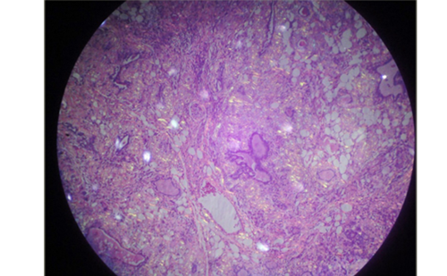
The presence of a diffuse goiter favors Graves’ disease, while the presence of a nodular goiter favors a toxic nodular goiter. Ophthalmopathy and pre-tibial myxedema are present only in Graves’ disease. A serum thyroid-stimulating immunoglobulin (TSI) antibody titer can be obtained to support the diagnosis of Graves’ disease, as 80–100% of untreated patients with Graves’ disease will have detectable TSH receptor antibody titers.18 A serum thyroid peroxidase antibody (TPO Ab) measurement may be helpful in distinguishing between autoimmune hyperthyroidism and gestational thyrotoxicosis. However, 5–10% of pregnant women without thyroid function abnormalities have detectable serum antithyroid antibody levels in the first trimester.19 One recent small study suggested that color flow Doppler sonography may be a useful adjunct in distinguishing Graves’ thyrotoxicosis from other causes of hyperthyroidism during pregnancy.20 Painless post-partum thyroiditis may occur following miscarriage, therapeutic abortions, or full-term deliveries, and therefore it occasionally occurs during pregnancy.21 In general, most pregnant women will tolerate mild thyrotoxicosis well, and repeating thyroid function tests in three to four weeks in cases where the etiology is uncertain is a reasonable approach.
In comparison, an iodine intake of 150μg/day is recommended for non-pregnant adults and adolescents.62
Treatment of Thyrotoxicosis in Pregnant Women
Women with untreated severe thyrotoxicosis during pregnancy are at increased risk for having low-birthweight infants, stillbirths, pre-term delivery, severe pre-eclampsia, congestive heart failure, placental abruption, and infants with congenital malformations, including premature closure of the fontanelles.22–24 For this reason, women of childbearing age who have been diagnosed with Graves’ disease should be advised to avoid pregnancy until euthyroidism has been achieved.Medical Therapy
The antithyroid drugs propylthiouracil (PTU) and methimazole (MMI) are the mainstay of therapy during pregnancy. Small amounts of PTU and MMI cross the placenta equally and may decrease fetal thyroid function and cause fetal goiter if given in excessive doses.25 Although most studies have not found clear dose–response relationships between antithyroid drug doses and the risk for neonatal thyroid dysfunction,26 treatment with relatively low doses of antithyroid drugs to keep the free T4 of pregnant women between the high to normal and slightly thyrotoxic range is recommended to reduce the risk for fetal hypothyroidism and goiter due to the transplacental passage of thionamides.27,28 Reassuringly, several studies have demonstrated that there are no cognitive delays in the children of mothers who took antithyroid medications during pregnancy.29 MMI has been described in occasional case reports as associated with cutis aplasia and esophageal and choanal atresia in newborns,30–32 although causal associations have not been proved. For these reasons, PTU is preferable to MMI for the treatment of hyperthyroidism in pregnancy.33 However, MMI is employed in countries where PTU is not available, and is used in the US in patients who are allergic to PTU, when begun later in pregnancy, and in non-compliant women, since MMI is effective in once-daily dosing compared with PTU, which is given three times daily.34 The frequent monitoring of thyroid function tests (usually every four weeks) is required throughout pregnancy in those taking antithyroid drugs. The dose of the antithyroid drug is often reduced as pregnancy progresses. Graves’ disease remits spontaneously in the last trimester of pregnancy in about 30% of women, and antithyroid drugs can be discontinued,35 but they often need to be re-started post-partum. Most beta blockers are rated pregnancy class C (risk cannot be ruled out; human studies are lacking) by the US Food and Drug Administration (FDA). Beta blockers can cross the placenta and have been associated with intrauterine growth restriction. However, the short-term use of beta blockers in pregnancy to control the symptoms of thyrotoxicosis is generally considered to be safe.17
Maternal and Fetal Monitoring
Graves’ disease frequently improves throughout gestation due to the immunosuppressive effects of pregnancy. Although easily transported across the placenta, titers of thyroid receptor-stimulating antibodies sufficient to cause fetal hyperthyroidism occur in only 1–5% of pregnancies complicated by maternal Graves’ disease.36 This may occur even in euthyroid pregnant women with Graves’ disease who have undergone thyroidectomy or radioactive iodine thyroid ablation prior to pregnancy. Thus, the thyroid receptor-stimulating antibody titer should be routinely measured in all patients with Graves’ disease between 26 and 28 weeks of pregnancy, a time when the placenta becomes more permeable to immunoglobulins.37 This can be accomplished using either a commercial TSI assay, in which values greater than 350–500% of controls raise concern for the development of fetal hyperthyroidism,38,39 or the thyrotropin-binding inhibiting immunoglobulin (TBII) assay, in which values greater than 40–70U/l suggest the possibility of fetal hyperthyroidism.40,41
A fetal ultrasound early in the third trimester should be considered to detect fetal goiter in women taking antithyroid medications and to assess for signs of fetal hyperthyroidism37 such as intrauterine growth retardation, goiter, and tachycardia.29,42 Large neonatal goiter due to either fetal Graves’ disease or antithyroid drugs that have crossed the placenta may cause airway obstruction after delivery.43 When a large fetal goiter is detected by ultrasound, resolution in utero has been reported with discontinuation of maternal PTU or MMI.44 In severe cases, injections of intra-amniotic L-thyroxine may be given,45 although this procedure entails a 0.5–1% risk of fetal complications.46
If suggested by ultrasound, fetal hyperthyroidism can be confirmed with umbilical blood sampling, although this is rarely necessary and confers some risk.47 In this situation, because antithyroid drugs cross the placenta, the mother could be treated with large PTU doses to achieve fetal thyroid hormone reduction, but would require the administration of L-thyroxine to maintain euthyroidism. The fetal status on this regimen can be monitored using serial ultrasounds with repeat cord blood sampling if absolutely necessary.47 Following delivery, infants with neonatal hyperthyroidism (in whom mortality may be as high as 16–25%) may have hyperkinesis, tremor, goiter, irritability, proptosis, excessive weight loss or failure to regain birthweight, premature closure of the fontanelles, and congestive heart failure starting in the first few days after birth and lasting for three to six months,41 when the infant’s blood TSI titer from the mother has dissipated. Severe neonatal or infantile hyperthyroidism may require treatment with antithyroid drugs, beta blockers, and, rarely, drugs that decrease the conversion of T4 to T3, such as dexamethasone. Finally, transient neonatal pituitary hypothyroidism following a hyperthyroid phase from uncontrolled maternal Graves’ disease has been described in Japan.48
Surgery
Thyroidectomy is only rarely required for the treatment of refractory hyperthyroidism, medication non-compliance, or when severe side effects of antithyroid drugs are present. When necessary, thyroidectomy is best performed in the second trimester.49
Hypothyroidism in PregnancyMaternal hypothyroidism, as characterized by an elevated TSH value, occurs in an estimated 2.5% of all US pregnancies.50 The fetal thyroid does not begin to concentrate iodine until weeks 10–12 of gestation and is not controlled by fetal pituitary TSH until approximately 20 weeks of gestation.51 Prior to this, the fetus is reliant on maternal T4, which crosses the placenta in very small quantities.52 Since thyroid hormone is required for normal neurodevelopment,53 even mildly low maternal T4 in pregnancy may result in cognitive delays in offspring,54,55 as may elevated maternal serum TSH values.5,56 However, one recent study found no relationship between isolated maternal hypothyroxinemia in early pregnancy and adverse perinatal events, such as low birthweight, low Apgar scores, requirement for intensive care unit monitoring, respiratory distress syndrome, major malformations, and neonatal death.57
Women with pre-existing hypothyroidism require larger amounts of L-thyroxine starting in the first trimester of pregnancy due to increased TBG levels and increased deiodination of T4 by the placenta. It has been recommended that L-thyroxine doses be increased by 30% as soon as pregnancy is confirmed, with close monitoring and dose adjustments thereafter as necessary.58 Ideally, a serum TSH level should be obtained as soon as pregnancy occurs. A conservative upper limit for TSH in the first trimester of pregnancy is 2.5mU/l, rising to 3.0mU/l for the second and third trimesters.8,59 Following delivery, L-thyroxine requirements typically decrease to pre-pregnancy levels and serum TSH levels should be monitored closely.60
Iodine Requirements in PregnancyThyroid hormone synthesis is dependent on adequate iodine intake, and an assurance of iodine sufficiency is especially important in pregnancy, when maternal thyroid hormone production normally increases by about 50%. The glomerular filtration rate of iodide also increases early in pregnancy, increasing renal iodide clearance and decreasing the circulating pool of plasma iodine.61 Due to the increased thyroid hormone production, increased renal iodine losses, and fetal iodine requirements in pregnancy, dietary iodine requirements are higher in pregnancy than they are for non-pregnant adults. Guidelines for daily dietary iodine intake during pregnancy are presented in Table 2 62,63,59. Due to concern about possible mild iodine deficiency among some US women of childbearing age, the American Thyroid Association (ATA) has recommended that all US and Canadian women receive dietary supplements containing 150µg iodine each day during pregnancy and lactation.64 Autoimmunity in Pregnancy and the Post-partum PeriodThyroid autoimmunity is common, with TPO Ab detectable in 13% of the US population,65 although the prevalence varies according to sex, age, and racial/ethnic group.66 The TPO antibody positivity is less frequent in African-American women than white women.67 Most TPO Ab-positive women do not have clinical hypothyroidism, although, as demonstrated by our recent data3 and others,2,68 they do tend to have higher serum TSH and lower free T4 values in the first trimester of pregnancy than women without detectable thyroid antibodies.
Relationship Between Thyroid Peroxidase Antibody Positivity and Obstetric Risks
The first report of increased miscarriage risk in association with antithyroid antibodies was published in 1990.69 Since then, multiple observational studies have demonstrated a two- to three-fold risk for m
scarriage among TPO Ab-positive women compared with TPO Ab-negative women, even in those with normal thyroid function.70–72 Some,73–75 but not all,76,77 studies have found associations between thyroid autoimmunity and increased rates of recurrent miscarriage. A two- to three-fold increase in the rate of miscarriage in women with TPO Ab undergoing assisted reproductive technology has been seen in four out of six studies performed to date.78–83 The presence of antithyroid antibodies has also been associated with a three-fold risk for premature delivery before 37 weeks of gestation.84 Another prospective study found a two-fold risk for very pre-term delivery (<32 weeks of gestation) in women with positive TPO Ab, but no increased risk for moderately pre-term delivery (32–37 weeks of gestation).85 Finally, the presence of TPO Ab in early pregnancy has also been associated with increased risk for post-partum thyroiditis.86
The reasons for the associations between antithyroid antibodies and risks for miscarriage and premature delivery remain unclear. They may be due to a direct effect of the antithyroid antibodies, or the antithyroid antibodies may serve as a marker for other, causative, autoimmune syndromes. Alternatively, antithyroid antibodies may simply indicate limited thyroid functional reserve.87 If TPO Ab-positive women are more likely to develop hypothyroidism during pregnancy, this hypothyroidism may be the cause of their obstetric complications.
Treatment of Pregnant Women with Thyroid Peroxidase Antibody Positivity
There has been one randomized clinical trial demonstrating that the treatment of euthyroid TPO Ab-positive women without a prior history of miscarriage could prevent adverse obstetric outcomes.84 The observations are most likely related to correction of mild thyroid hypofunction in the treated women, since L-thyroxine therapy would not be expected to eliminate underlying autoimmunity. This study was performed in Italy, an area of mild to moderate dietary iodine deficiency, so the generalizability to women in the US is limited and the study has not yet been replicated.
Maternal–Fetal Medicine
Painless Post-partum Thyroiditis
Painless post-partum thyroiditis, an autoimmune disorder, causes lymphocytic inflammation of the thyroid within the first few months after delivery in up to 10% of US women.88,89 It is more common in women with high serum TPO Ab concentrations in the first trimester of pregnancy or immediately post-partum. It is also more common in women with other autoimmune disorders, such as type 1 diabetes, or with a family history of autoimmune thyroid disease.An inflammatory thyrotoxicosis caused by leakage of pre-formed thyroid hormone typically begins one to six months post-partum and lasts for one to two months. A hypothyroid phase may follow, starting four to eight months post-partum and lasting for four to six months. Only one-third of patients will experience the classic triphasic thyroid hormone pattern. While 80% of women will recover normal thyroid function within one year, 50% became permanently hypothyroid within seven years in one study.90 After one episode of post-partum thyroiditis, there is a 70% chance of recurrence with subsequent pregnancies.91
A small, non-tender, firm goiter is usually present in post-partum thyroiditis. High serum TPO and/or thyroglobulin antibody concentrations are present in most cases.92 The 24-hour radioactive iodine uptake (RAIU) test may be used to distinguish post-partum thyroiditis from post-partum Graves’ disease, as uptake will be low (<5%) rather than elevated as in Graves’ disease. The RAIU test should be performed in patients with symptomatic thyrotoxicosis when no clear signs of Graves’ disease such as large goiter or ophthalmopathy are present. As radioactive iodine is secreted in breast milk and 123I has a half-life of 13 hours, nursing mothers need to pump and discard milk for at least two days after the test. Since serum TSI is present in most cases of Graves’ disease and usually absent in painless post-partum thyroiditis, serum TSI level is also helpful in differentiating between the two disorders. The thyrotoxic phase of post-partum thyroiditis rarely requires therapy, but when severe can be treated with beta-blockers. Antithyroid drug therapy is contraindicated because excess thyroid hormone synthesis does not occur. Treatment of the hypothyroid phase may not be necessary, but if prolonged or the patient is symptomatic, L-T4 should be given and withdrawn after six to nine months in order to determine whether thyroid function has normalized. These women should be screened yearly for thyroid dysfunction in view of the reported occurrence of hypothyroidism years later.90
Thyroid Screening in Pregnancy—Current Clinical Guidelines
The question of whether or not all pregnant women should be screened with thyroid function tests and/or tests for antithyroid antibodies is extremely controversial. The American Association of Clinical Endocrinologists (AACE) has recommended routine TSH screening before pregnancy or during the first trimester in all pregnant women.93 The American College of Obstetricians and Gynecologists (ACOG) has recommended against serum TSH testing in asymptomatic pregnant women.17,51 Recent guidelines from the Endocrine Society recommend that pregnant women undergo aggressive case-finding, but not screening.59 However, case-finding in pregnant women has limitations: it has been reported that screening pregnant women with a family or personal history of thyroid disease or other autoimmune disease would fail to identify 30% of women with overt or subclinical hypothyroidism.94 Given the potential obstetric and neonatal complications of untreated thyroid disorders in pregnancy, we recommend the ascertainment of maternal serum thyroid function as early as possible in the gestational course.