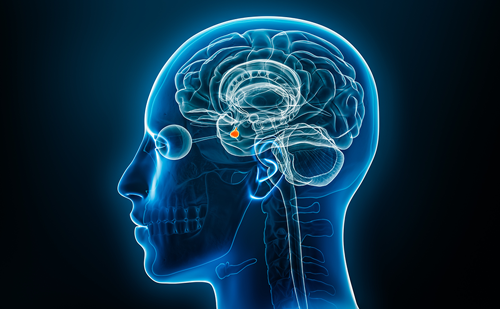
Adult-onset growth hormone deficiency (AO-GHD) is most often caused by pituitary or hypothalamic tumours or their treatment, and may serve as a model where the effect of chronic GH deficiency on skeletal metabolism can be studied. While the low bone mass in adults with childhood-onset GHD (CO-GHD) may be explained by deficient bone accretion during childhood, decreased bone mass in AO-GHD may be caused by imbalanced bone remodelling.
Adult-onset growth hormone deficiency (AO-GHD) is most often caused by pituitary or hypothalamic tumours or their treatment, and may serve as a model where the effect of chronic GH deficiency on skeletal metabolism can be studied. While the low bone mass in adults with childhood-onset GHD (CO-GHD) may be explained by deficient bone accretion during childhood, decreased bone mass in AO-GHD may be caused by imbalanced bone remodelling. These patients have secondary osteoporosis characterised by reduced bone mass, decreased bone turnover measured by biochemical markers and increased fracture risk. However, studies on the impact of GH substitution have yielded conflicting results, probably due to high doses and short treatment periods. Longer studies, with treatment periods of one year or more, have shown significant increases in bone mass and turnover.
GH plays a crucial role in the maintenance of bone mass in adults by regulating bone remodelling through a complex interaction of circulating GH, insulin-like growth factors (IGFs) and IGF-binding proteins (IGFBPs) and locally produced IGFs and IGFBPs acting in an autocrine and paracrine way. The cellular basis for these interactions has been thoroughly studied, employing in vitro systems with isolated homogenous bone cell populations, and the molecular signalling pathways revealed. Furthermore, progress in genetic engineering has greatly increased the understanding of how GH controls somatic growth in vivo. The original somatomedin hypothesis originated in the 1950s in an effort to describe how somatic growth was regulated by the pituitary and that the effects of GH on target tissue were mediated by intermediate substances and not GH alone.1
At present, it appears clear that GH also may have direct effects on target tissues and that locally produced IGF-1 may mediate the effects of GH. The debate now largely concerns the importance of liver-derived IGF-1.2 This article focuses on recent work on the effect of GH/IGF on remodelling in patients with AO-GHD and experimental models characterised by decreased systemic levels of these proteins.
There are other papers that provide detailed reviews on GH and IGF signalling in vitro and the somatomedin hypothesis.3 The effects of GH and IGF-1/-2 on bone cells in vitro are briefly described in Figure 1.
Studies in Genetically Altered Animals
The GH receptor (GHR) and IGF type 1 receptor (IGF1R) are present in many tissues and various systemic factors may regulate local expression of IGFs and IGFBPs in the intact organism. The use of genetically altered mice has had a major impact on defining the role of IGFs in skeletal homeostasis, and especially the role of systemic IGF-1 in the development and maintenance of the adult skeleton. Studies in mice lacking GHR demonstrate reduced cortical and longitudinal bone growth, decreased bone turnover and a markedly reduced bone mineral content (BMC).4,5 Many of these effects can be substantially reversed by IGF-1 treatment, suggesting that the main defect may relate to reduced IGF-1 levels in the absence of GHR.4
Mice rendered deficient in IGF-1 show reduced bone size as expected; however, trabecular bone (TB) volume is markedly increased, especially in female mice, due to increased connectivity, increased number and decreased spacing of the trabeculae. This indicates that the actions of IGF-1 on bone are sexually dimorphic and suggests an interaction between sex steroid hormones and IGF-1 in these actions.6 Thus, lack of IGF-1 leads to the development of a bone structure that, although smaller, appears more compact, possibly due to decreased IGF-1-mediated bone resorption or increased responsiveness to GH.6
Liver-specific knockouts with decreased systemic IGF-1 levels show that liver-derived IGF-1 exerts a small but significant effect on cortical bone growth, while it is not required for the maintenance of TB in adult mice.7,8 However, double gene disruption of acid-labile subunit and IGF leads to a further decline in IGF-1 and a significant decrease in bone mineral density (BMD), suggesting that a threshold level of circulating IGF-1 may be necessary to maintain bone mass.8 Still, these animals have increased circulating GH levels, which could explain the maintained TB through direct effects on osteoblasts. Finally, IGF-1 messenger (m)RNA levels are unchanged in bone in these mice, indicating that local IGF-1 production is enough to maintain TB volume.7 Accordingly, osteoblast-specific knockout of IGF1R decreases TB volume,9 while mice with overexpression of IGF-1 targeted to osteoblasts have increased TB volume.10 It should be mentioned that IGF-1 knockouts may also display 1,25-dihydroxyvitamin D deficiency and elevated parathyroid hormone (PTH) levels.11
Thus, the net effects of GH and IGF-1 on bone structure are complex, region- and bone-specific and influenced by other hormones, not least sex steroids. GH may have direct effects on osteoblasts and may increase bone volume. Liver-derived IGF-1 may be of importance for cortical bone, but does not seem to be required for the maintenance of the TB in adult mice.
Studies in Patients with AO-GHD
Patients with GHD have secondary osteoporosis characterised by reduced bone mass,12–15 decreased bone turnover as measured by biochemical markers and increased fracture risk.15–17 Notably, a recent study, including both CO-GHD and AO-GHD, indicated the effect of severe GHD on BMD at several sites to be partly age-dependent, with BMD z scores above the reference population in elderly patients, and a significantly higher BMD compared with young GHD adults, suggesting a protective effect of low bone turnover in relation to the age-related bone loss.18 Although GHD patients have many other pituitary deficiencies that may impact bone metabolism, epidemiological studies have revealed that GHD alone explains the increased fracture risk associated with these patients.15,17
Treatment of GHD patients with GH dose-dependently increases bone turnover as judged by biochemical bone markers.19–24 Due to the dynamics of bone remodelling (bone resorption preceding bone formation), increases in bone resorptive and formative markers are observed after three and six months substitution, respectively. Although the effects of GH on bone turnover are consistent and sustained during long-term substitution, the effects on bone mass have been more elusive due to short duration of treatment period.19,20,22,23,25 In fact, earlier studies with a treatment period of up to one year demonstrated decreased BMD, probably due to increased remodelling activity with increased remodelling space and a larger proportion of new unmineralised bone. This might partly be explained by the use of unphysiological high GH doses not taking gender into account. Longer studies, with treatment periods of two years or more physiological doses, have shown significant increases in bone mass.20,22,26–32 BMD continues to rise long after cessation of GH replacement, suggesting that this hormone initiates the bone remodelling process but is not required to sustain such an effect.33,34 Thus far, long-term randomised studies on AO-GHD patients treated with individual doses of GH aiming at normalising IGF-1 have not been published. Previous studies have either been open or used a fixed or weight-related dosing. If individualised, the regime has initially been based on a high standard dose titrated down according to IGF-1 levels.35
These changes in bone mass are positively correlated with increases in serum IGFBPs, as well as GH and IGF-1, suggesting that GH may increase bone mass partly through changes in systemic levels of IGF family members.36 Furthermore, enhanced cortical bone protein and gene expression of IGF-1 is found during GH therapy in patients with AO-GHD, substantiating that the effects of GH may be mediated by enhanced local production of IGFs, secondary to increased systemic levels.37,38 This is in accordance with observations in cortical but not trabecular bone from acromegalic patients.39,40 Moreover, these changes are correlated with changes in bone matrix gene expression of the calcitonin receptor as well as biochemical bone markers, indicating a direct effect of locally produced IGF-1 on osteoclasts and in regulating bone turnover.38 Additional treatment with alendronate in GHD patients receiving stable GH replacement therapy is effective in further increasing BMD at the lumbar spine.41 Also, treatment of GHD adults with IGF-1 increases bone formation without increasing bone resorption, suggesting that IGF-1 may exert a direct anabolic effect on bone forming cells in vivo and that local increases in IGF-1 gene and protein expression are secondary to effects of GH on osteoblasts.42 Similar effects are found in GH-deficient transgenic mice treated with IGF-1.43
The effects of GH on BMD seem to be gender-dependent, with greater effects of GH substitution in men than women.27,29,31 Although the precise mechanisms underlying these differences are unclear, it seems likely that sex steroids may play a role. Physiological oestrogen replacement therapy in GHD women leads to a relative resistance to the stimulatory effect of GH on IGF-1 production.44 Also, there may be an antagonism between oestrogen and GH at the peripheral tissue level.45 Furthermore, patients with AO-GHD may have reduced sensitivity to the effects of PTH on kidney and bone. While GH replacement increases PTH target organ sensitivity, this effect is reduced and delayed in women, leading to a delayed increase in bone turnover markers following GH therapy.46
The Osteoprotegerin–Receptor Activator of NFkappaB–Receptor Activator of NFkappaB Ligand Axis
IGF-1 may act as one of several coupling agents by activating bone formation and bone resorption. Thus, the amount of IGF-1 released from bone matrix should activate a proportionate response from osteoblasts to produce enough osteoid to fill the resorption lacunae. In addition to direct effects on osteoclasts, GH and IGF-1 may affect bone resorption indirectly by stimulating release of paracrine mediators that regulate osteoclastic bone resorption. Critical for this process is the balance between the newly discovered members of the tumour necrosis factor ligand and receptor superfamilies, osteoprotegerin (OPG) and receptor activator of NFkappaB ligand (RANKL), which mediate the effects of many upstream regulators of bone metabolism.47
By binding its receptor, RANK, RANKL stimulates osteoclast differentiation, activates mature osteoclasts, and inhibits osteoclast apoptosis, as shown in vitro, and is a sufficient and necessary factor for osteoclast formation and, thus, bone resorption.48–50 OPG blocks the effects of RANKL by neutralising and preventing binding to its receptor RANK.
Age-related changes in OPG have been observed in both serum and bone matrix,51,52 suggesting that OPG may be regulated by age-related factors such as GH/IGF-1. Possibly, the increase in serum OPG found in metabolic bone disease may be compensatory to increased osteoclastic bone resorption. Still, OPG does not seem to be a marker of bone turnover as serum OPG levels were normal in patients with acromegaly, as well as GHD.53 Furthermore, no changes in serum OPG were seen during GH substitution to AO-GHD women or elderly.38,54
Another study found increased serum OPG following GH substitution to a mixed population of patients with GHD, negatively correlated to changes in bone turnover.55 In contrast, Rubin et al. found that IGF-1 increased RANKL and decreased OPG expression in mouse stromal cells, favouring pro-resorptive activity in vitro.54 Still, serum levels may not necessarily reflect the cytokine levels in the bone microenvironment, and in vitro models may not account for other OPG-regulating cytokines influenced by GH/IGF-1. Thus, increased OPG protein and gene expression has been demonstrated in cortical bone explants following GH substitution, reflecting the in vivo situation locally in bone. Nonetheless, increased cortical OPG expression may protect against IGF-1-induced bone resorption and potentially be of importance for the long-term beneficial effects of GH replacement. Further studies investigating the OPG/RANKL system in transgenic GH/IGF models may clarify these issues.
Conclusions
Bone mass and turnover is reduced in AO-GHD, leading to clinically significant osteoporosis with increased vertebral fracture rate. Treatment with GH increases bone turnover and long-term intervention leads to increased bone mass. Still, long-term clinical studies on AO-GHD patients treated individually in order to normalise IGF-1 are missing. However, studies in genetically altered animals indicate that the net effect of GH and IGF-1 on bone structure are, as mentioned above, complex, region and bone-specific and influenced by other hormones, including sex steroids. Studies on IGFBP regulation of IGF action and effects GH and the IGF family on osteoclastic resorption, and in particular the OPG–RANK–RANKL axis, may help to clarify the role of GH/IGF-1 in these patients and lead to more effective therapeutic modalities. ■