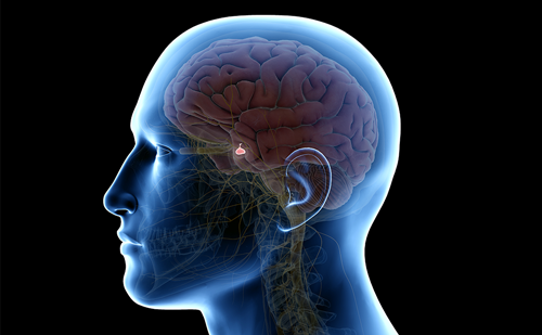
Prolactin (PRL) is a peptide hormone secreted by lactotroph cells of the anterior pituitary. PRL is predominantly under inhibitory influence of hypothalamic dopamine, whereas thyrotropin-releasing hormone (TRH), among other endogenous substances, is the main PRL-releasing factor. In physiological conditions, PRL secretion is also affected by numerous exogenous influences, such as stress, physical effort, hypoglycaemia, lactation and sleep.
Prolactin (PRL) is a peptide hormone secreted by lactotroph cells of the anterior pituitary. PRL is predominantly under inhibitory influence of hypothalamic dopamine, whereas thyrotropin-releasing hormone (TRH), among other endogenous substances, is the main PRL-releasing factor. In physiological conditions, PRL secretion is also affected by numerous exogenous influences, such as stress, physical effort, hypoglycaemia, lactation and sleep.
The main activity of PRL is its influence on the mammary gland (the so-called mammotrophic and lactotrophic effect) and the gonads, inhibiting the pulsatile excretion of gonadotropin-releasing hormone (GnRH) and the secretion of follicle-stimulating hormone (FSH) and luteinising hormone (LH).
Since human PRL was purified in 1972, the clinical syndrome of hyperprolactinaemia has been characterised extensively, the predominant symptoms being galactorrhoea, oligomenorrhoea or secondary amenorrhoea, infertility in women and reduced libido, impotence and gynaecomastia in men. In children and adolescents, major clinical signs of hyperprolactinaemic syndrome are delayed puberty, gynaecomastia, galactorrhoea and primary amenorrhoea.
Hyperprolactinaemia represents a common diagnostic dilemma due to the fact that it is often encountered in clinical practice even in the absence of significant pathology. An elevated PRL value, also in asymptomatic patients, creates the need to rule out diverse organic and functional aetiologies of hyperprolactinaemia (see Table 1). Therefore, differential diagnosis of hyperprolactinaemia may be difficult and undiscounted – especially in children and adolescents, in whom the stress of venipuncture even causes mild PRL increase.
Prolactinomas
Prolactinomas, the most common functioning pituitary tumours, are the most frequent cause of hyperprolactinaemia in patients aged two to 80 years; however, these tumours are less frequent in children. Also, hyperprolactinaemia may derive from a pituitary tumour that contains bipotential cells secreting PRL and another pituitary hormone, e.g. growth hormone (GH), or from two distinct adenomas producing various hormones. From this point of view, these hyperprolactinaemic syndromes may overlap other endocrine conditions with affected pituitary function.
For pituitary prolactinoma, the clinical syndrome traditionally described in children is caused by functional alterations resulting from hyperprolactinaemia (i.e. galactorrhoea, gynaecomastia, amenorrhoea and delayed puberty) and/or by the mass effect of the tumour (i.e. headaches, visual field abnormalities, etc.). Interestingly, there is a predominance of macroprolactinoma over microprolactinoma in most paediatric studies; at the time of diagnosis, males present mainly with neurophthalmologic abnormalities secondary to tumoural growth, whereas in females, endocrine-gynaecological symptoms are predominant.
The absolute PRL levels have considerable diagnostic and predictive value because the serum PRL concentrations are consistently elevated in patients with prolactinoma. For many years, it has been argued that PRL values above 100ng/ml are suggestive of organic pathology, while values below that level are indicative of functional alteration. The most likely cause of a PRL level above 250ng/ml is a PRL-secreting tumour, whereas tumours causing compression of the pituitary stalk show only moderate elevation of PRL resulting from an insufficient dopamine supply. In children and adolescents with prolactinoma, serum PRL levels are 10 times higher in macroadenoma than in microadenoma. Indeed, a PRL concentration of more than 600ng/ml is always due to a macroprolactinoma. However, many patients affected by prolactinoma have PRL levels below 100ng/ml in the first measurement.
The PRL response to TRH is characteristically blunted in patients with prolactinoma, but the response may be normal. In addition, prolactinoma patients with normal basal PRL levels may have PRL values that are more than three times elevated after stimulation. Finally, the usefulness of dynamic tests of the PRL axis is highly controversial.
Misdiagnosis
On the other hand, not all hyperprolactinaemic patients with uncertain pituitary imaging have an adenoma. Peillon et al. reported a pseudotumoural appearance of the pituitary on computed tomography (CT) or magnetic resonance imaging (MRI) scans in five women with functional hyperprolactinaemia. In these cases, the tissue pathology showed lactotroph hyperplasia, which is also found in physiological conditions such as pregnancy or lactation. It is important to recognise that functioning hyperprolactinaemia may mimic PRL-secreting adenoma on CT and MRI scans. Follow-up CT and MRI brain scans at regular intervals (initially at one year, then every two years thereafter) are necessary to monitor for the development of pituitary tumour.
Furthermore, all hyperprolactinaemic syndromes may be misdiagnosed as prolactinoma due to the relatively common occurrence of pituitary incidentaloma on imaging studies. A tiny pituitary mass revealed by MRI scan is likely to be an incidentaloma of little clinical significance. CT and MRI imaging in the general population, undertaken for reasons other than suspected pituitary disease, indicate that 10–20% of such images are consistent with the presence of a pituitary adenoma. Given that up to 10% of a population may harbour an asymptomatic adenoma, it is to be expected that some patients will be identified who have both hyperprolactinaemia and imaging findings consistent with a pituitary adenoma. It has been suggested that this may account for part of the failure of pituitary microsurgery of prolactinoma. Hyperprolactinaemia is frequently reported in patients with inherited endocrine syndromes, such as Carney complex, McCune-Albright syndrome (MAS) or multiple endocrine neoplasia type 1 (MEN 1), which share some endocrine and skin features. The vast majority of hyperprolactinaemic patients with MAS or Carney complex have negative pituitary gland imaging studies, whereas prolactinomas are commonly present in kindreds with MEN 1. Therefore, hyperprolactinaemia in MEN 1, contrary to MAS and Carney complex, is a clinically distinct entity due to pituitary adenoma and constitutive activation of somatomammotroph cells, respectively. It is always prudent to exclude these inherited endocrine syndromes for all hyperprolactinaemic patients.
Polycystic Ovary Syndrome
Hyperprolactinaemia is the cause of 20–30% of secondary amenorrhoea, a condition that affects at least 3% of women in reproductive age, and polycystic ovary syndrome (PCOS) is diagnosed in 80% of patients with menstrual cycle disturbances. Indeed, PCOS causes such as stress, hypoglycaemia and obesity are reported as the main causes of hyperprolactinaemia.
PCOS is a chronic hyperandrogenous disease with characteristic androgenic phenotype, disturbances of the menstrual cycle, hirsutism and reduced hormonal carbohydrate and lipid metabolism. There are also reports of hyperinsulinism and insulin resistance in PCOS. Hyperinsulinaemia is a reason for hyperprolactinaemia in PCOS as well as in obesity; hyperinsulinism may induce hyperandrogenaemia through inhibition of androgen catabolism. Also, oestrogens result from the aromatase conversion of androgen excess. It should be noted that, among the numerous factors stimulating PRL secretion, oestrogens play a significant role, which is the case in PCOS or obesity or after oral intake of oestrogens. Hyperoestrogenism may stimulate PRL release by lactotrophs directly as well as inhibiting dopamine secretion in the hypothalamus. Significantly higher levels of PRL have been found in PCOS than in control females with regular cycles and menstruation; the highest PRL values are detected in women with both PCOS and obesity. As the PCOS diagnosis is straightforward and pharmacological treatment leads to restoration of pituitary function, it is mandatory to rule out PCOS in all young hyperprolactinaemic females.
Other Aetiologies of Hyperprolactinaemia
Rarely, marginal increases in PRL levels accompany other endocrine conditions, such as hypothyroidism. Due to the fact that TRH, among other endogenous substances, is the main PRL-releasing factor, increased PRL secretion may be due to primary hypothyroidism. Therefore, complete thyroid function must be evaluated in all hyperprolactinaemic patients because thyroid hormone replacement rapidly normalises PRL function in these patients.
Hyperprolactinaemia also results from conditions that decrease dopamine action at the lactotroph cells, such as pregnancy, seizures or drugs, and conditions in which reduced clearance of PRL occurs, such as chronic renal failure. Many pharmacological treatments may also raise pituitary PRL release, such as dopamine receptor antagonists, atypical antipsychotic agents, antidepressants, oestrogens, opiates and antihypertensive drugs. Furthermore, reversible asymptomatic hyperprolactinaemia is reported as an adverse effect during GnRH agonist (GnRHa) treatment, such as triptorelin, in girls. Interestingly, hyperprolactinaemic GnRHa-treated children show PRL levels that are elevated by more than three times after TRH stimulation. However, increased PRL values rapidly normalise after GnRHa withdrawal.
Transient increase in PRL levels has been confirmed during and following seizures in both adults and children. For PRL levels to increase postictally, neuromuscular activity lasting for a minimum of 30 seconds is required. Febrile seizures are characterised by their brief duration and benign, non-epileptic nature; thus, the transient postictal PRL elevation of epileptic seizures may not be obligatory. In addition to the postictal hyperprolactinaemia, it has been proposed that patients with epilepsy may have high interictal PRL levels, but this has not been confirmed worldwide. While high postictal PRL levels may indicate a convulsive attack, a normal PRL value does not guarantee its absence. Therefore, once drug effects or seizure episodes have been excluded, it is mandatory to check renal function and pregnancy status in hyperprolactinaemic subjects.
Ruling out Macroprolactinaemia
Finally, for all patients found to have hyperprolactinaemia, it is prudent to exclude macroprolactinaemia. There are three major molecular forms of PRL in the circulation:
- monomeric PRL (little PRL; 23 kilo Daltons (kDa));
- dimeric PRL (or big PRL; 45–50kDa); and
- macroprolactin (big-big PRL, 150–170kDa).
Macroprolactin is a complex of immunoglobulin G (IgG) and monomeric PRL with prolonged plasma half-life due to delayed renal clearance. PRL mainly circulates in the monomeric form (85–95%), but occasionally, macroprolactin may be the predominant form, such as in macroprolactinaemia. The estimated prevalence of macroprolactin among patients with hyperprolactinaemia ranges from 9% to 42%, and its prevalence in healthy people is less than 1%. Although biological activity has been demonstrated in vitro, it is generally believed the macroprolactin lacks biological activity in vivo because it cannot cross the endothelial lining and reach the cell surface receptors. Therefore, macroprolactinaemia – the presence of elevated levels of PRL of high molecular mass with little, if any, bioactivity – is one aetiology of hyperprolactinaemia that is considered benign.
Unfortunately, macroprolactin is a significant source of analytical error that is affecting most, if not all, of the PRL assays in current use. Recent studies have indicated that macroprolactinaemia accounts for up to 26% of biochemical hyperprolactinaemia depending on the immunoassay in use and, thus, macroprolactinaemia represents a common diagnostic pitfall that is responsible for frequent misdiagnosis and mismanagement of hyperprolactinaemic patients. It is advisable for clinicians to have some knowledge of the assay proprieties used by their laboratories. One of the commonly used methods to screen for macroprolactinaemia is polyethylene glycol (PEG) precipitation. PRL that is not precipitated by PEG is considered as free and biologically active PRL. Knowing the level of monomeric PRL is desirable for hyperprolactinaemic patient management as this is the biologically active form, and monomeric PRL and macroprolactin can occur simultaneously. However, despite convincing evidence of the importance of ruling out macroprolactinaemia in hyperprolactinaemic subjects, routine screening has not yet been generally adopted.
Summary
In conclusion, although hyperprolactinaemia represents a common problem encountered in clinical practice, its diagnosis may be confused or overlooked. Therefore, an accurate differential diagnosis is mandatory in order to rule out the diverse aetiologies of hyperprolactinaemia. In addition, although dopamine agonist drugs, such as bromocriptine, are often efficacious at reducing PRL levels in all hyperprolactinaemic syndromes, both method and duration of curative treatment largely depends on the specific aetiology of the hyperprolactinaemia. Failing to make a differential diagnosis, the hyperprolactinaemic condition could lead to unnecessary and costly investigations, inappropriate intervention and needless apprehension. ■
A version of this article, with references, can be found in the Reference Section on the website supporting this business briefing (www.touchbriefings.com).


