touchENDOCRINOLOGY touchENDOCRINOLOGY
Diabetes
Read Time: 12 mins

Epigenetics of the Pathogenesis and Complications of Type 2 Diabetes Mellitus

Copy Link
Published Online: Apr 14th 2023 touchREVIEWS in Endocrinology. 2023:46-53 DOI: https://doi.org/10.17925/EE.2023.19.1.46
Authors: Velmurugan Mannar, Hiya Boro, Deepika Patel, Sourabh Agstam, Mazhar Dalvi, Vikash Bundela
Quick Links:
Abstract
Article
Article Information
Abstract:
Overview

Epigenetics of type 2 diabetes mellitus (T2DM) has widened our knowledge of various aspects of the disease. The aim of this review is to summarize the important epigenetic changes implicated in the disease risks, pathogenesis, complications and the evolution of therapeutics in our current understanding of T2DM. Studies published in the past 15 years, from 2007 to 2022, from three primary platforms namely PubMed, Google Scholar and Science Direct were included. Studies were searched using the primary term type 2 diabetes and epigenetics with additional terms such as ‘risks’, ‘pathogenesis’, ‘complications of diabetes’ and ‘therapeutics’. Epigenetics plays an important role in the transmission of T2DM from one generation to another. Epigenetic changes are also implicated in the two basic pathogenic components of T2DM, namely insulin resistance and impaired insulin secretion. Hyperglycaemia-induced permanent epigenetic modifications of the expression of DNA are responsible for the phenomenon of metabolic memory. Epigenetics influences the development of micro and macrovascular complications of T2DM. They can also be used as biomarkers in the prediction of these complications. Epigenetics has expanded our understanding of the action of existing drugs such as metformin, and has led to the development of newer targets to prevent vascular complications. Epigenetic changes are involved in almost all aspects of T2DM, from risks, pathogenesis and complications, to the development of newer therapeutic targets.

Keywords

DNA methylation, epigenetics, histone modification, metabolic memory, microRNA, type 2 diabetes mellitus

Article:

Article highlights

  • Epigenetics refers to the heritable changes in DNA expression without changes in the genetic code.

  • Epigenetic changes are brought about by post-translational modifications of histone proteins, covalent modifications of DNA bases and microRNA.

  • Epigenetics explains how environmental milieu such as diet, physical activity, circadian rhythm, intrauterine malnutrition or maternal obesity interact with the genome of an individual and lead to diseases such as type 2 diabetes mellitus (T2DM).

  • Epigenetics also contributes substantially to the development of micro and macrovascular complications of T2DM.

  • Current research to develop newer drugs that target the epigenetic dysregulation in T2DM is ongoing.

Diabetes has become a global pandemic, with an estimated 536.6 million people living with diabetes worldwide in 2021, and this is likely to increase to 783.2 million by the year 2045.1 The primary pathophysiology of type 2 diabetes mellitus (T2DM) involves insulin resistance in the liver, adipose tissue and skeletal muscle, followed by defects in insulin secretion later in the course of the disease.2 Notably, T2DM is a polygenic disorder that develops complex interactions between genes and the environment.

Epigenetics refers to heritable changes in DNA expression without alterations in the genetic code.3 In the past few decades, understanding the epigenetics of T2DM has unravelled the missing pathogenic links in the causation of the disease.4 Simultaneously, it has also enabled us to understand the effects of environmental factors, such as diet and physical activity, on the pathogenesis of the disease. Epigenetic changes can be used as potential biomarkers to assess the risk of the onset of T2DM and vascular complications. Epigenetic alterations can also predict the response to therapy and lifestyle interventions, thereby offering a tool for precision medicine.4

With the ever-increasing knowledge of epigenomics,5 it is not possible to review all epigenetic mechanisms of T2DM comprehensively. In this review, we discuss the basic mechanisms of epigenetics and try to summarize the important epigenetic dysregulation implicated in pathogenesis, vascular complications and therapeutics to appreciate its impact on our current understanding of T2DM.

Basics of pigenetic mechanisms

In the 1940s, the term epigenetics was used to refer to the complex interactions between the genome and the environment.6 This concept has evolved significantly in the past 50 years. Riggs et al. defined epigenetics as the study of mitotically and/or meiotically heritable changes in gene function that cannot be explained by changes in the DNA sequence.3 Epigenetics-mediated differential gene expression can explain the heterogeneous functions of different cell types in the body despite them all carrying the same genetic information.6 It is now known that epigenetic changes also control noncoding regions of DNA, which are essential in various physiological and pathophysiological states.7 Epigenetic changes can be classified into two types: direct epigenetics and indirect epigenetic, which is further subdivided into within indirect epigenetics and across indirect epigenetics.8 Direct epigenetics refers to the changes in gene expression occurring in an individual’s lifespan due to interactions with the environment. Within indirect epigenetics refers to the changes in gene expression that occur within the intrauterine environmentFinally, across indirect epigenetics refers to altered gene expression due to epigenetic changes inherited from ancestors.8 Hence, epigenetic changes can influence an individual’s genomic expression from when they are in the zygote state and throughout their whole lifespan, both as a static expression (i.e. inherited from their ancestors) and a dynamic expression (i.e. due to interactions with the environment).

The process of protein synthesis takes place in two steps: transcriptionwhich is the synthesis of messenger RNA (mRNA) by copying a gene’s DNA sequence, followed by translation, in which the information carried by mRNA is decoded to produce peptides by ribosomal RNA and transfer RNA. Epigenetics-mediated altered DNA expression can occur at the level of both transcription and translation. Covalent modifications of DNA bases (methylation) and modifications of histone proteins alter DNA expression at the level of transcription, whereas noncoding RNAs (specifically microRNAs [miRNAs]) affect the gene expression at the level of translation (Figure 1).8,9

Figure 1: Mechanism of epigenetic changes
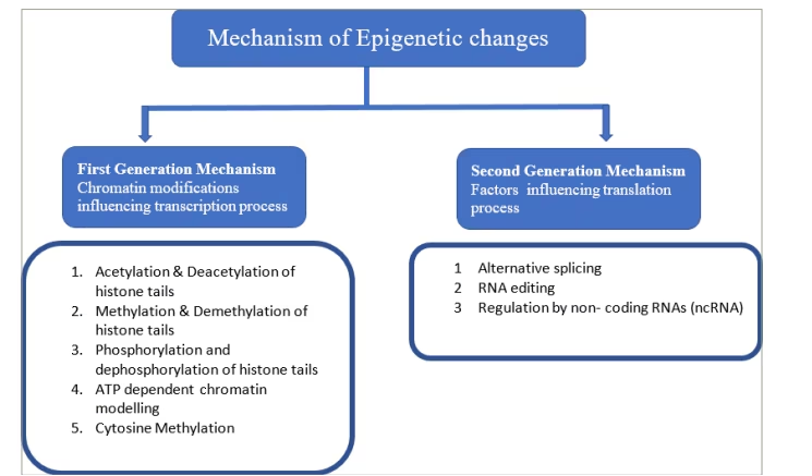
ATP = adenosine triphosphate.

DNA methylation is an enzymatic process by which methyl group is covalently added to cytosine residues to alter the gene expression. It is carried out by enzymes belonging to the families of DNA methyl transferases (DNMT), namely DNMT1, DNMT2, DNMT3A, DNMT3B and DNMT3L.8–10 S-adenosyl methionine (methyl donor) donates methyl groups to cytosine residues in CpG, or cytosine guanine (CG), dinucleotide sites. In humans, most of the promoter sites of DNA have a CpG island that is the target of these enzymes. The hypermethylation of the CpG sites of these target promoters prevents the process of transcription, thus silencing gene expression (Figure 2).11

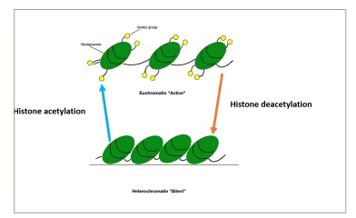
Histone proteins can be modified by methylation, acetylation, ubiquitination or phosphorylation.12 These modifications lead to changes in the structure of chromatin, causing the formation of either euchromatin, which results in gene expression, or heterochromatin, which results in gene silencing.13 Histone methylation can lead to the activation or repression of gene expression. Histone acetylation leads to a decrease in the positive charges of histone proteins, which in turn causes a decrease in its interactions with DNA and increased accessibility of transcription complexes to DNA (euchromatin), leading to increased gene expression. The opposite happens when deacetylation takes place (Figure 3).13

Figure 2: Methylated and unmethylated CpG islands of DNA causing gene repression and expression, respectively
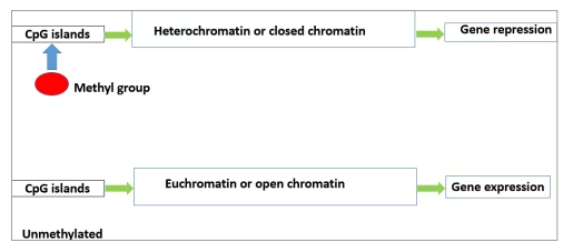
Figure 3: Histone acetylation causing the formation of euchromatin (active chromatin) and deacetylation causing the formation of heterochromatin (silent chromatin)
miRNAs are specialized noncoding RNAs about the size of 22 nucleotides in length.14 They are synthesized by DNA with the help of RNA polymerase II. From the nucleus, they reach the cytoplasm to bind to specific target mRNA, resulting in the cleavage of bound mRNA causing translational repression (Figure 4).14 MiRNAs can be regulated by processes of DNA methylation, RNA modification and histone modification.

Figure 4: Mechanism of microRNA causing cleavage of target messenger RNA, leading to translational repression
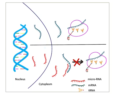
miRNA = microRNA; mRNA = messenger RNA; tRNA = transfer RNA.

Epigenetics and type 2 diabetes mellitus

Epigenetics and risk of type 2 diabetes mellitus

The role of epigenetics in the onset of T2DM can be exemplified by studies showing an increased prevalence of diabetes in adulthood when there is a history of poor maternal nutrition or diabetes in the mother during gestation.15,16 It should be noted that both maternal malnutrition and gestational diabetes put the child at future risk of developing T2DM. Creating an undernutritional environment in utero, especially in early gestation, was hypothesized to lead to the epigenetic programming of various metabolic pathways in the foetus in anticipation of an adverse environment later in life.16 When these children are postnatally exposed to an abundance of nutrition, they are at risk of developing T2DM, obesity and metabolic syndrome. This phenomenon is referred to as the thrifty phenotype hypothesis. It has been proposed that maternal malnutrition leads to decreased levels of leptin in the blood, which may later lead to obesity.16 Studies of the Dutch Hunger Winter famine cohort found that children exposed to famine in early gestation showed a 5.2% decrease in DNA methylation of the insulin-like growth factor 2 (IGF2) gene differentially methylated region compared with children who had normal maternal nutrition in pregnancy or were exposed to famine during the later part of pregnancy.17 Apart from these methylation changes, increased expression of miR-5765p seems to play an important role in the pathogenesis of increased cardiometabolic risk.17 Furthermore, lower circulating levels of miR-15a, miR-29b, miR-126 and miR-223 and higher levels of miR-283p can predict the risk of developing T2DM.18

During postnatal life, factors such as diet, physical activity, sleepwake cycle and various environmental factors lead to changes in the epigenome and contribute to the risk of developing T2DM in the future.15 An important RNA-binding protein, NONO, is an epigenetic regulator of genes, controlling various pathways of carbohydrate and fat metabolism in the liver in accordance with the availability of nutrition.19 NONO is pivotal in predicting an individual’s risk of developing T2DM.15,19

Yajnik depicted the thin-fat Indian phenotype in an article where he describes that Indian babies are born with low birth weight but are found to have higher visceral fat than their English counterparts and are predisposed to insulin resistance and T2DM in later life.20 The Developmental Origin of Health and Disease theory traces the origin of T2DM to intrauterine life followed by rapid childhood growth leading to a biphasic nutritional insult.21 Yajnik et al. have also reported that nutritional factors such as 1C (methyl) metabolism with normal to high maternal folate levels and vitamin B12 deficiency in mothers predisposed Indian babies to higher adiposity and insulin resistance by foetal epigenetic changes.22

The complex interactions of epigenetics in the evolution and progression of T2DM are summarized in Figure 5.15

Figure 5: Epigenetics and type 2 diabetes – complex interactions at various levels
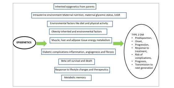
IUGR = intrauterine growth restriction; T2DM = type 2 diabetes mellitus.

The epigenetics and pathogenesis of type 2 diabetes mellitus

Two primary components in the pathogenesis of T2DM are insulin resistance and impaired insulin secretion. Various studies have demonstrated altered DNA methylation of genes, namely PPARGKCNQ1TCF7L2 and insulin receptor substrate 1 (IRS1)that are involved in the actions of insulin at sites such as the liver, skeletal muscle and adipose tissue.23–31 Pancreatic islets of patients with T2DM have shown decreased expression of genes involved in insulin secretion, such as PPARGC1AINS and PDX1, due to increased methylation.32–34 Decreased expression of PPARGC1A is also noted in the skeletal muscles of subjects with T2DM.35 Further human and animal studies of skeletal muscle suggested methylation defects in genes that regulate insulin sensitivity, such as NDUFB6,36 COX5a,37 OXPHOS,38 PGC-1α,39 PDK4 and PPAR-δ.40 Studies in rats with obesity induced by a high-fat diet suggested increased methylation and, subsequently, decreased expression of glucokinase and L-type pyruvate kinase promoter regions, which are involved in the pathogenesis of insulin resistance in the liver.41 Upregulation of histone deacetylase 7 (HDAC7) in human pancreatic cells of individuals with T2DM was found to be associated with a decrease in glucose-mediated insulin secretion.42 Acetylation of the FOXO1 gene, which controls PDX1, leads to an impact on beta-cell development and glucose homeostasis.43 HDAC6-mediated histone3 lysine 9 (H3K9) deacetylation leads to the downregulation of the IRS2 protein, which in turn leads to the development of insulin resistance.44 MiRNAs were found to be important in the processes of betacell dysfunction and betacell survival, both of which are crucial events in the pathogenesis of T2DM.45 The miRNA MiR375 participates in the development of the pancreas and decreases insulin secretion by inhibiting myotrophin.46 Another miRNA, miR124a, impacts insulin secretion with a mechanism similar to that of miR-375.47 MiR-29a and MiR-29b inhibit the secretion of insulin by their inhibitory actions on monocarboxylate transporter 1 (MCT1).48 MiR-184 can induce betacell replication, thus causing an increase in the betacell population.49 MiRNAs also contribute to the pathogenesis of insulin resistance by their action on the phosphoinositol-3 kinase (PI3K)/AKT and other insulin signalling pathways.50 The following MiRNAs regulate these proteins: miR-128a, miR-96 and miR-126, which control the expression of the IRS-1; miR-29, miR-3845p and miR-1, which regulate PI3K expression; miR-143, miR-145, miR-29, miR-383, miR-33a, miR-33b and miR-21, which modulate AKT expression; and miR-133a, miR-133b, miR-223 and miR-143, which control the expression of the glucose transporter GLUT4.51

Epigenetics and complications of type 2 diabetes mellitus

Metabolic memory

Metabolic memory, or legacy effect, refers to the benefits of early good glycaemic control to the overall positive effects in the course of T2DM.52,53 Good glycaemic control early in the natural history of T2DM leads to long-term protection from micro and macrovascular complications irrespective of the glycaemic status in the later part of the disease.52,53 On the other hand, due to metabolic memory, initial poor glycaemic control may lead to a higher risk for vascular complications in the later course of the disease.52 The evidence for this effect comes from three large, randomized controlled trials with long-term follow-up data, namely the Diabetes control and complications trial in type 1 diabetes,54 the United Kingdom prospective diabetes study55 and Steno-2 in T2DM.56 In these studies, patients who were in the initial intensive arm continued to show decreased incidence of vascular (both micro and macro) complications on long-term follow-up compared with patients who were initially in the conventional arm and later switched to intensive therapy despite presenting similar glycaemic status in the later phases.

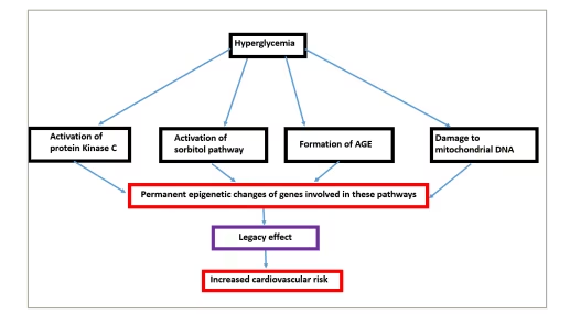
The basic pathophysiological mechanisms of metabolic memory are hyperglycaemia-induced damage to mitochondrial DNA and its proteins, stimulation of protein kinase C, activation of sorbitol pathway and formation of advanced glycation end products.57 It has been proposed that hyperglycaemia leads to permanent epigenetic modifications of DNA expression of the abovementioned pathways that are consistent with poor glycaemic states, and they continue to persist even after good control is achieved at a later stage (Figure 6).58 Epigenetic mechanisms include changes in post-translational histone modifications, DNA methylation and miRNAs leading to irreversible changes, referred to as metabolic memory.59

Figure 6: Mechanism of legacy effect

AGE = advanced gycation end product.

Endothelial dysfunction

Endothelial dysfunction seems to be a crucial component in the development of all vascular complications of diabetes. Chronic, uncontrolled hyperglycaemia leads to vascular damage by multiple pathways, namely oxidative stress, increased production of advanced glycation end products, activation of inflammatory and fibrotic pathways by transforming growth factor-beta (TGF-β), nuclear factor κβ (NF-κβ) and angiotensin II (AngII).15 Endothelin 1 (ET-1)a peptide produced by the vascular endothelium, causes vasoconstriction and increased fibrosis, and is known to be abundant in patients with vascular complications in T2DM.60 Methylation defects are noted in the CpG regions of the promoter of the EDN1 gene, which leads to its overexpression.59 Decreased methylation and increased acetylation of histone proteins in the NF-κB gene promoter region leads to overexpression of this proinflammatory marker in the endothelial cells.61 Increased acetylation of histone H3K9/K14 leads to the overexpression of other inflammatory markers, such as interlukin-8 (IL8) and heme oxygenase gene 1 (HMOX1), in the endothelial cells of the aorta.61 Hyperglycaemia affects H3K9 demethylation, leading to increased expression of matrix metallopeptidase-9 (MMP-9), which in turn causes damage to the mitochondria and endothelial cell death.62 MiR-1405p, miR-2213p, miR-200b and miR-130b-3p participate in the pathogenesis of endothelial dysfunction by targeting several genes related to apoptosis, inflammation, hyperpermeability, senescence and pathological angiogenesis.63 Apart from the these microRNAs, miR-126 overexpression is associated with endothelial dysfunction of peripheral arterial disease in T2DM.64

Macrovascular complications

DNA methylation defects are noted in genes involved in the formation of atherosclerotic plaques, such as SOD2, FGF2, ABCA1, COX2 and SMAD 7.65 DNA demethylation causing overexpression of the KLF4KLF5 and OPN genes leads to the increased mitotic activity of smooth muscle cells in coronary vasculature.66 Hyperacetylation of histone proteins H3K9 and H3K27 has been implicated in stabilizing atherosclerotic plaques.67 Moreover, alteration in histone proteins associated with oxidized low-density lipoproteinmediated inflammatory response is crucial in the pathogenesis of coronary artery disease (CAD) in T2DM. In one study, miR-126 levels were found to be inversely correlated with serum low-density lipoprotein in T2DM patients with underlying CAD compared with T2DM without CAD.68 Two important miRNAs, namely miR-1 and miR-133, have been shown to be significantly correlated to the risk for CAD in T2DM, such that they can be considered biomarkers for macrovascular complications.69,70 Other miRNAs, such as miR-210,71 miR-2172 and miR-370,73 are also shown to be associated with T2DM-related CAD. Altered levels of miR-451a,74 miR-1955p75 and miR-146a76 are associated with cerebrovascular events in T2DM.

Microvascular complications

Diabetic retinopathy

Diabetic retinopathy (DR) is an important cause of blindness in adults worldwide and its presentation spectrum can range from nonproliferative retinoapthy (mild, moderate and severe) to proliferative retinopathy, which is associated with new vessel formation and haemorrhages. Both proliferative and nonproliferative DR can be associated with macular oedema, which can further cause severe morbidity. Global DNA methylation seems to be increased early in the course of the development of DR but does not increase further with disease progression.77,78 Animal studies have demonstrated the contribution of hypermethylation of mitochondrial DNA and DNA polymerase gamma to the pathogenesis of DR.79–81 The modification of histone proteins seems to be associated with neuronal cell death and increased vascular permeability of retinal vessels.82,83 Methylation of the histone protein H3K9 by the histone methyl transferase encoded by SUV39H2 is associated with DR onset.84 Increased H3 histone acetylation is noted in animal models of DR linked to activation of the NF-kβ inflammatory pathway.85 In uncontrolled T2DM, alterations in histone proteins lead to MMP-9 overexpression in retinal capillaries, leading to mitochondrial dysfunction and cell death.86 Wide arrays of miRNAs take part in the initiation and progression of DR. MiR-126 regulates the expression of vascular endothelial growth factor 1 (VEGF1) and other vascular adhesion molecules, which are of considerable importance to the pathogenesis of proliferative DR.87 There is decreased expression of miR-31 and miR-184, which in physiological states inhibit new vessel formation.88 Increased expression of miR-21 is of paramount importance in the pathogenesis of DR by contributing to endothelial dysfunction.89 Circulating miRNAs can be used as biomarkers for the early and late complications of DR. One example of such miRNA is miR-210, the levels of which are higher in individuals with DR than in those without.90 Furthermore, the levels of miR-210 are much higher in proliferative DR than in non-proliferative DR.

Diabetic nephropathy

Diabetesrelated kidney disease is the leading cause of chronic kidney disease worldwide and can present both with or without albuminuria. Genome-wide studies have shown that increased DNA methylation is correlated with inflammation in diabetic nephropathy.91,92 In animal models of T2DM nephropathy, hypermethylation of the promoter region of Ras protein activator like 1 (RASAL1) has been noted.93,94 Increased expression of transforming growth factor-beta 1 (TGFβ1) in diabetic kidney disease leads to hypermethylation of RASAL-1 causing activation of Ras-GTP signalling.95 This mechanism results in collagen deposition and fibrosis, which is an important step in the pathogenesis of diabetic nephropathy96,97 Altered cytosine methylation in the promotor regions of the mammalian target of rapamycin (mTOR) is conducive to the inflammation of nephropathy.95 Another study showed decreased expression of the transcription factor Krüppel-like factor 4 (KLF4), leading to the hypermethylation of the nephrotic syndrome type 1 (NPHS1) gene, which encodes nephrin, podocyte cell death and albuminuria.98,99 Decreased methylation of the myoinositol oxygenase (MIOX) gene is associated with diabetic nephropathy progression by increasing oxidative stress and fibrosis.100 Alteration of histone proteins leads to increased TXNIP gene expression, causing increased inflammation of mesangial cells.101 High levels of expression of HDAC4 in diabetic nephropathy promote inflammatory changes by inhibiting the STAT1 pathway.102 Mouse models of streptozotocin-induced albuminuria have shown reduced expression of Sirtuin 1/silent information regulator 1 (SIRT1)histone deacetylase, which leads to decreased expression of claudin1 in podocytes.103,104 MiR-133b, miR-199b,105 miR-23a106 and miR-30e107 contribute to renal fibrosis, whereas miR-146a is associated with the activation of inflammatory pathways.108 Furthermore, the urinary exosomal miRNA can potentially be used as a biomarker for the onset of diabetic nephropathy.109 For example, increased urinary levels of miR-19155p,110 miR-8773p,111 miR-192 and miR-215 are found in patients with T2DM with diabetic nephropathy.112

Diabetic neuropathy

Diabetic neuropathy is the most common microvascular complication of diabetes, is due to involvement of long nerve fibres and usually presents as distal symmetric polyneuropathy. Decreased DNA methylation of the whole genomic DNA in white blood cells is a potential biomarker for diabetic neuropathy.113 DNA methylation defects have been seen across many genes coding for proteins affecting multiple steps in the pathogenesis of diabetic neuropathy, such as axon guidance, glycerophospholipid metabolism and mitogen-activated protein kinase (MAPK) signalling pathways.114 Hypermethylation of the NINJ2 gene and its subsequent decreased expression have been observed in diabetic neuropathy.115 This protein, expressed in Schwann cells, is essential for the regeneration of peripheral nerves after injury.115 Increased expression of miR-199a3p, which causes the downregulation of the extracellular serine protease inhibitor E2 (Serpin E2), has been attested in the peripheral blood of patients with diabetic neuropathy.116 Decreased expression of miR-25,117 miR-146118,119 and miR-190a5p120 has been noted in animal models of diabetic neuropathy. This results in the modulation of oxidative stress, interleukins and various other inflammatory processes resulting in neuronal injury.121 Other miRNAs significant to the pathogenesis of diabetic neuropathy are miR-128a, miR-155a and miR-499a, which can potentially be used as biomarkers for the screening of diabetic neuropathy.122

Diabetic cardiomyopathy

Diabetic cardiomyopathy is defined as presence of cardiac dysfunction in patients with diabetes in the absence of any other explainable causes such as CAD, valvular heart disease or hypertenion. Epigenetic changes lead to the overexpression of genes of the reninangiotensinaldosterone system (RAAS) axis, which is crucial in the pathogenesis of diabetic cardiomyopathy.123–125 Diabetic cardiomyopathy is associated with hypermethylation of the protein sarcoplasmic/endoplasmic reticulum calcium-ATPase 2a (SERCA2a), which is physiologically important for cardiac muscle relaxation.126 This decreased expression of SERCA2a can explain the diastolic dysfunction seen in diabetic cardiomyopathy.123 MiRNAs are pivotal in various steps of the pathogenesis of cardiomyopathy, such as muscle hypertrophy,127 fibrosis,128 mitochondrial dysfunction,129 cell death130 and foetal genetic programming.131 Foetal genetic reprogramming involves decreased expression of the alpha myosin heavy chain (α-MHC) gene and increased expression of the beta myosin heavy chain (β-MHCgene, which contribute to the development of diabetic cardiomyopathy.131 Altered expression of some miRNAs – namely miR-1,132 miR-146a,133 miR-133a,134 miR-150,135 miR-200c,136 miR-1523p,137 miR-26a/b-5p,138 miR-29b-3p139 and miR-223140 – is crucial in the hypertrophy and fibrosis of cardiac muscles.

Epigenetics and therapeutics of type 2 diabetes mellitus

Understanding epigenetics has led to the possibility of using epigenetic changes as biomarkers for predictjng the risk of developing T2DM, its complications and for the development of therapeutic targets. As discussed above, many miRNA levels can be used as biomarkers of microvascular and macrovascular complications. There is evidence to suggest that inhibition of specific miRNAs that are involved in the loss of betacell function or betacell death can result in the improvement of betacell functions.45 Histone deacetylase inhibitors can improve insulin sensitivity by increasing the acetylation of lysine amino acids in the insulin receptor substrate 2 (IRS2) protein.141 Apabetalone, a new drug, acts by blocking histone interactions with DNA; this action has been shown to prevent the rise in inflammatory proteins and the development of atherosclerotic plaques.142 Supplementation with lactobacillus can cause changes in the histone methylation profile and improve insulin resistance.143 Treatment of T2DM with metformin causes reduced DNA methylation of genes coding for metfomin transporters, leading to their increased expression and, thus upregulating the beneficial effects on glycaemic control and insulin resistance.144,145 Metformin also causes alteration in the expression of various histone methyl transferases and SIRT1 (deacetylase).144 Glucagon-like peptide 1 (GLP1) receptor analogues, which are currently used for managing T2DM, help preserve betacell function by histone modifications and reactivation of pdx-1 transcription.146,147 GLP1 receptor analogues also lead to improvements in the fatty liver, which is mediated by modulation of SIRT1 (deacetylase) and decrease in the expression of NF‐κB.148

Newonset diabetes that occurs during statin therapy is postulated to be due to DNA methylation defects leading to the dysregulation of ATP-binding cassette subfamily G member 1 (ABCG1).149

MiRNA-based therapeutics aimed at targeting various levels of the pathogenesis of T2DM are being testein animal studies.150–154 MiRNA inhibitors can be used to suppress the overexpression of pathogenic miRNA, and miRNA mimics can be used for overcoming the underexpression pathology.155,156 Antisense oligonucleotides can also be used to modulate miRNA expression.

Conclusions

This review summarizes the evidence supporting the substantial contribution of epigenetics to the pathogenesis and complications of T2DM. Epigenetic modifications start in the intrauterine environment and continue throughout an individual’s life. Epigenetics influences the transmission of T2DM across generations. It also explains how adverse environmental milieus such as food habits, sedentary lifestyle, circadian rhythm, maternal malnutrition or maternal obesity interact with an individual’s genome, leading to various disease states such as T2DM. In addition, epigenetics is instrumental in developing various micro and macrovascular complications of T2DM. Research is being conducted to develop epigenetic biomarkers that predict the risk of T2DM and its vascular complications. Newer drugs under development aim to correct the epigenetic dysregulation in T2DM. However, further research is required to identify the epigenetic regulators specific to T2DM before novel therapies addressing the pathogenesis and complications of T2DM can be developed.

Article Information:
Disclosure

Velmurugan Mannar, Hiya Boro, Deepika Patel, Sourabh Agstam, Mazhar Dalvi and Vikash Bundela have no financial or non-financial relationships or activities to declare in relation to this article.

Compliance With Ethics

This article involves a review of the literature and did not involve any studies with human or animal subjects performed by any of the authors.

Review Process

Double-blind peer review.

Authorship

The named authors meet the International Committee of Medical Journal Editors (ICMJE) criteria for authorship of this manuscript, take responsibility for the integrity of the work as a whole, and have given final approval for the version to be published.

Correspondence

Hiya Boro, Department of Endocrinology and Metabolism, Aadhar Health Institute, House No 1913, Sector 16- 17, Hisar, Haryana 125001, India. E: hiya21288@gmail.com

Support

No funding was received in the publication of this article.

Access

This article is freely accessible at touchENDOCRINOLOGY.com. © Touch Medical Media 2023

Data Availability

Data sharing is not applicable to this article as no datasets were generated or analysed during the writing of this article.

Received

2022-12-22

References

1. Sun HSaeedi PKaruranga Set alIDF diabetes atlas: Global, regional and country-level diabetes prevalence estimates for 2021 and projections for 2045Diabetes Res Clin Pract2022;183:109119DOI10.1016/j.diabres.2021.109119

2. Ali OGenetics of type 2 diabetesWorld J Diabetes2013;4:11423DOI10.4239/wjd.v4.i4.114

3. Riggs ADPorter TNOverview of epigenetic mechanismsInRussoVEAMartienssenRRiggsAD, eds. Epigenetic Mechanisms of Gene RegulationCold Spring Harbor, NYCold Spring Harbor Laboratory Press19962945.

4. Ling CBacos KRönn TEpigenetics of type 2 diabetes mellitus and weight change – A tool for precision medicine? Nat Rev Endocrinol2022;18:43348DOI10.1038/s41574-022-00671-w

5.National Cancer InstituteEpigenomics and epigenetics researchAvailable athttps://epi.grants.cancer.gov/epigen/#:~:text=Epigenetics%20focuses%20on%20processes%20that,a%20cell%20or%20entire%20organism (accessed date27 March 2023)

6. Waddington CHThe epigenotypeEndeavour1942;1:1820.

7. Zhou ZLin ZPang Xet alEpigenetic regulation of long non-coding RNAs in gastric cancerOncotarget2018;9:1944358DOI10.18632/oncotarget.23821

8. Lacal IVentura REpigenetic inheritance: Concepts, mechanisms and perspectivesFront Mol Neurosci2018;11:292DOI10.3389/fnmol.2018.00292

9. Dupont CArmant DRBrenner CAEpigenetics: Definition, mechanisms and clinical perspectiveSemin Reprod Med2009;27:35157DOI10.1055/s-0029-1237423

10. Jin BLi YRobertson KDDNA methylation: Superior or subordinate in the epigenetic hierarchy? Genes Cancer2011;2:60717DOI10.1177/1947601910393957

11. Schaefer MPollex THanna Ket alRNA methylation by DNMT2 protects transfer RNAs against stress-induced cleavageGenes Dev2010;24:159095DOI10.1101/gad.586710

12. Zarkesh MEhsandar SHedayati MGenetic and epigenetic aspects of type 2 diabetes mellitus: A reviewAustin Endocrinol Diabetes Case Rep2016;1:1004.

13. Małodobra-Mazur MCierzniak AMyszczyszyn Aet alHistone modifications influence the insulin-signaling genes and are related to insulin resistance in human adipocytesInt J Biochem Cell Biol2021;137:106031DOI10.1016/j.biocel.2021.106031

14. O’Brien JHayder HZayed YPeng COverview of microRNA biogenesis, mechanisms of actions, and circulationFront Endocrinol (Lausanne)2018;9:402DOI10.3389/fendo.2018.00402

15. Dhawan SNatarajan REpigenetics and type 2 diabetes riskCurr Diab Rep2019;19:47DOI10.1007/s11892-019-1168-8

16. Smith CJRyckman KKEpigenetic and developmental influences on the risk of obesity, diabetes, and metabolic syndromeDiabetes Metab Syndr Obes2015;8:295302DOI10.2147/DMSO.S61296

17. Doan TNAAkison LKBianco-Miotto TEpigenetic mechanisms responsible for the transgenerational inheritance of intrauterine growth restriction phenotypesFront Endocrinol (Lausanne)2022;13:838737DOI10.3389/fendo.2022.838737

18. Chen HLan HYRoukos DHCho WCApplication of microRNAs in diabetes mellitusJ Endocrinol2014;222:R1R10DOI10.1530/JOE-13-0544

19. Benegiamo GBrown SAPanda SRNA dynamics in the control of circadian rhythmAdv Exp Med Biol2016;907:10722DOI10.1007/978-3-319-29073-7_5

20. Yajnik CSConfessions of a thin-fat IndianEur J Clin Nutr2018;72:46973DOI10.1038/s41430-017-0036-3

21. Bianco-Miotto TCraig JMGasser YPet alEpigenetics and DOHAD: From basics to birth and beyondJ Dev Orig Health Dis2017;8:51319DOI10.1017/S2040174417000733

22. Yajnik CSDeshpande SSJackson AAet alVitamin B12 and folate concentrations during pregnancy and insulin resistance in the offspring: The Pune Maternal Nutrition StudyDiabetologia2008;51:2938DOI10.1007/s00125-007-0793-y

23. Barrès ROsler MEYan Jet alNon-CPG methylation of the PGC-1alpha promoter through DNMT3B controls mitochondrial densityCell Metab2009;10:18998DOI10.1016/j.cmet.2009.07.011

24. Nilsson EJansson PAPerfilyev Aet alAltered DNA methylation and differential expression of genes influencing metabolism and inflammation in adipose tissue from subjects with type 2 diabetesDiabetes2014;63:296276DOI10.2337/db13-1459

25. Nilsson EMatte APerfilyev Aet alEpigenetic alterations in human liver from subjects with type 2 diabetes in parallel with reduced folate levelsJ Clin Endocrinol Metab2015;100:E1491501DOI10.1210/jc.2015-3204

26. Nitert MDDayeh TVolkov Pet alImpact of an exercise intervention on DNA methylation in skeletal muscle from first-degree relatives of patients with type 2 diabetesDiabetes2012;61:332232DOI10.2337/db11-1653

27. Ribel-Madsen RFraga MFJacobsen Set alGenome-wide analysis of DNA methylation differences in muscle and fat from monozygotic twins discordant for type 2 diabetesPLoS One2012;7:e51302DOI10.1371/journal.pone.0051302

28. Kirchner HSinha IGao Het alAltered DNA methylation of glycolytic and lipogenic genes in liver from obese and type 2 diabetic patientsMol Metab2016;5:17183DOI10.1016/j.molmet.2015.12.004

29. Abderrahmani AYengo LCaiazzo Ret alIncreased hepatic PDGF-AA signaling mediates liver insulin resistance in obesity-associated type 2 diabetesDiabetes2018;67:131021DOI10.2337/db17-1539

30. Baumeier CSaussenthaler SKammel Aet alHepatic DPP4 DNA methylation associates with fatty liverDiabetes2017;66:2535DOI10.2337/db15-1716

31. You DNilsson ETenen DEet alDnmt3a is an epigenetic mediator of adipose insulin resistanceElife2017;6:120DOI10.7554/eLife.30766

32. Yang BTDayeh TAKirkpatrick CLet alInsulin promoter DNA methylation correlates negatively with insulin gene expression and positively with hba(1c) levels in human pancreatic isletsDiabetologia2011;54:3607DOI10.1007/s00125-010-1967-6

33. Ling CDel Guerra SLupi Ret alEpigenetic regulation of PPARGC1A in human type 2 diabetic islets and effect on insulin secretionDiabetologia2008;51:61522DOI10.1007/s00125-007-0916-5

34. Yang BTDayeh TAVolkov PAet alIncreased DNA methylation and decreased expression of PDX-1 in pancreatic islets from patients with type 2 diabetesMol Endocrinol2012;26:120312DOI10.1210/me.2012-1004

35. Gillberg LJacobsen SCRibel-Madsen Ret alDoes DNA methylation of PPARGC1A influence insulin action in first degree relatives of patients with type 2 diabetes? PLoS One2013;8:e58384DOI10.1371/journal.pone.0058384

36. Ling CPoulsen PSimonsson Set alGenetic and epigenetic factors are associated with expression of respiratory chain component NDUFB6 in human skeletal muscleJ Clin Invest2007;117:342735DOI10.1172/JCI30938

37. Gong YLiu YLi Jet alHypermethylation of cox5a promoter is associated with mitochondrial dysfunction in skeletal muscle of high fat diet-induced insulin resistant ratsPLoS One2014;9:e113784DOI10.1371/journal.pone.0113784

38. Rönn TPoulsen PHansson Oet alAge influences DNA methylation and gene expression of COX7A1 in human skeletal muscleDiabetologia2008;51:115968DOI10.1007/s00125-008-1018-8

39. Barres RKirchner HRasmussen Met alWeight loss after gastric bypass surgery in human obesity remodels promoter methylationCell Rep2013;3:10207DOI10.1016/j.celrep.2013.03.018

40. Barrès RYan JEgan Bet alAcute exercise remodels promoter methylation in human skeletal muscleCell Metab2012;15:40511DOI10.1016/j.cmet.2012.01.001

41. Jiang MZhang YLiu Met alHypermethylation of hepatic glucokinase and L-type pyruvate kinase promoters in high-fat diet-induced obese ratsEndocrinology2011;152:12849DOI10.1210/en.2010-1162

42. Daneshpajooh MBacos KBysani Met alHDAC7 is overexpressed in human diabetic islets and impairs insulin secretion in rat islets and clonal beta cellsDiabetologia2017;60:11625DOI10.1007/s00125-016-4113-2

43. Nakae JBiggs WHKitamura Tet alRegulation of insulin action and pancreatic beta-cell function by mutated alleles of the gene encoding forkhead transcription factor FOXO1Nat Genet2002;32:24553DOI10.1038/ng890

44. Dalfrà MGBurlina SDel Vescovo GGLapolla AGenetics and epigenetics: New insight on gestational diabetes mellitusFront Endocrinol (Lausanne)2020;11:602477DOI10.3389/fendo.2020.602477

45. Ofori JKKaragiannopoulos ANagao Met alHuman islet microRNA-200c is elevated in type 2 diabetes and targets the transcription factor ETV5 to reduce insulin secretionDiabetes2022;71:27584DOI10.2337/db21-0077

46. Dumortier OFabris GPisani DFet alMicroRNA-375 regulates glucose metabolism-related signaling for insulin secretionJ Endocrinol2020;244:189200DOI10.1530/JOE-19-0180

47. Sebastiani GPo AMiele Eet alMicroRNA-124a is hyperexpressed in type 2 diabetic human pancreatic islets and negatively regulates insulin secretionActa Diabetol2015;52:52330DOI10.1007/s00592-014-0675-y

48. Pullen TJda Silva Xavier GKelsey GRutter GAMiR-29a and mir-29b contribute to pancreatic beta-cell-specific silencing of monocarboxylate transporter 1 (mct1)Mol Cell Biol2011;31:318294DOI10.1128/MCB.01433-10

49. Tattikota SGRathjen THausser Jet alMiR-184 regulates pancreatic β-cell function according to glucose metabolismJ Biol Chem2015;290:2028494DOI10.1074/jbc.M115.658625

50. Zhong F-YLi JWang Y-Met alMicroRNA-506 modulates insulin resistance in human adipocytes by targeting S6K1 and altering the IRS1/PI3K/AKT insulin signaling pathwayJ Bioenerg Biomembr2021;53:67992DOI10.1007/s10863-021-09923-2

51. Improta-Caria ACDe Sousa RALRoever Let alMicroRNAs in type 2 diabetes mellitus: Potential role of physical exerciseRev Cardiovasc Med2022;23:29DOI10.31083/j.rcm2301029

52. Aschner PJRuiz AJMetabolic memory for vascular disease in diabetesDiabetes Technol Ther2012;14(Suppl. 1):S6874DOI10.1089/dia.2012.0012

53. Zhang LChen BTang LMetabolic memory: Mechanisms and implications for diabetic retinopathyDiabetes Res Clin Pract2012;96:28693DOI10.1016/j.diabres.2011.12.006

54. Nathan DMCleary PABacklund J-Yet alIntensive diabetes treatment and cardiovascular disease in patients with type 1 diabetesN Engl J Med2005;353:264353DOI10.1056/NEJMoa052187

55. Holman RRPaul SKBethel MAet al10-year follow-up of intensive glucose control in type 2 diabetesN Engl J Med2008;359:157789DOI10.1056/NEJMoa0806470

56. Gæde POellgaard JCarstensen Bet alYears of life gained by multifactorial intervention in patients with type 2 diabetes mellitus and microalbuminuria: 21 years follow-up on the steno-2 randomised trialDiabetologia2016;59:2298307DOI10.1007/s00125-016-4065-6

57. Testa RBonfigli ARPrattichizzo Fet alThe “metabolic memory” theory and the early treatment of hyperglycemia in prevention of diabetic complicationsNutrients2017;9:437DOI10.3390/nu9050437

58. Reddy MAZhang ENatarajan REpigenetic mechanisms in diabetic complications and metabolic memoryDiabetologia2015;58:44355DOI10.1007/s00125-014-3462-y

59. Jin JWang XZhi XMeng DEpigenetic regulation in diabetic vascular complicationsJ Mol Endocrinol2019;63:R103115DOI10.1530/JME-19-0170

60. Ergul AEndothelin-1 and diabetic complications: Focus on the vasculaturePharmacol Res2011;63:47782DOI10.1016/j.phrs.2011.01.012

61. Prattichizzo FGiuliani ACeka Aet alEpigenetic mechanisms of endothelial dysfunction in type 2 diabetesClin Epigenetics2015;7:56DOI10.1186/s13148-015-0090-4

62. Kowluru RAShan YRole of oxidative stress in epigenetic modification of MMP-9 promoter in the development of diabetic retinopathyGraefes Arch Clin Exp Ophthalmol2017;255:95562DOI10.1007/s00417-017-3594-0

63. Kowluru RAShan YMishra MDynamic DNA methylation of matrix metalloproteinase-9 in the development of diabetic retinopathyLab Invest2016;96:10409DOI10.1038/labinvest.2016.78

64. Zampetaki AKiechl SDrozdov Iet alPlasma microrna profiling reveals loss of endothelial mir-126 and other micrornas in type 2 diabetesCirc Res2010;107:8107DOI10.1161/CIRCRESAHA.110.226357

65. Pang MLi YGu Wet alRecent advances in epigenetics of macrovascular complications in diabetes mellitusHeart Lung Circ2021;30:18696DOI10.1016/j.hlc.2020.07.015

66. Prandi FRLecis DIlluminato Fet alEpigenetic modifications and non-coding RNA in diabetes-mellitus-induced coronary artery disease: Pathophysiological link and new therapeutic frontiersInt J Mol Sci2022;23:4589DOI10.3390/ijms23094589

67. Greißel ACulmes MBurgkart Ret alHistone acetylation and methylation significantly change with severity of atherosclerosis in human carotid plaquesCardiovasc Pathol2016;25:7986DOI10.1016/j.carpath.2015.11.001

68. Al-Kafaji GAl-Mahroos GAbdulla Al-Muhtaresh Het alCirculating endothelium-enriched microRNA-126 as a potential biomarker for coronary artery disease in type 2 diabetes mellitus patientsBiomarkers2017;22:26878DOI10.1080/1354750X.2016.1204004

69. Liu NBezprozvannaya SWilliams AHet alMicroRNA-133a regulates cardiomyocyte proliferation and suppresses smooth muscle gene expression in the heartGenes Dev2008;22:324254DOI10.1101/gad.1738708

70. Ikeda SHe AKong SWet alMicroRNA-1 negatively regulates expression of the hypertrophy-associated calmodulin and mef2a genesMol Cell Biol2009;29:2193204DOI10.1128/MCB.01222-08

71. Amr KSAbdelmawgoud HAli ZYet alPotential value of circulating microRNA-126 and microRNA-210 as biomarkers for type 2 diabetes with coronary artery diseaseBr J Biomed Sci2018;75:827DOI10.1080/09674845.2017.1402404

72. Al-Hayali MASozer VDurmus Set alClinical value of circulating microribonucleic acids mir-1 and mir-21 in evaluating the diagnosis of acute heart failure in asymptomatic type 2 diabetic patientsBiomolecules2019;9:193DOI10.3390/biom9050193

73. Liu HYang NFei Zet alAnalysis of plasma mir-208a and mir-370 expression levels for early diagnosis of coronary artery diseaseBiomed Rep2016;5:3326DOI10.3892/br.2016.726

74. Li PTeng FGao Fet alIdentification of circulating microRNAs as potential biomarkers for detecting acute ischemic strokeCell Mol Neurobiol2015;35:43347DOI10.1007/s10571-014-0139-5

75. Giordano MTrotta MCCiarambino Tet alCirculating miRNA-195-5p and -451a in diabetic patients with transient and acute ischemic stroke in the emergency departmentInt J Mol Sci2020;21:7615DOI10.3390/ijms21207615

76. Ortega FJMercader JMMoreno-Navarrete JMet alProfiling of circulating microRNAs reveals common microRNAs linked to type 2 diabetes that change with insulin sensitizationDiabetes Care2014;37:137583DOI10.2337/dc13-1847

77. Lee RWong TYSabanayagam CEpidemiology of diabetic retinopathy, diabetic macular edema and related vision lossEye Vis (Lond)2015;2:17DOI10.1186/s40662-015-0026-2

78. Maghbooli ZHossein-nezhad ALarijani Bet alGlobal DNA methylation as a possible biomarker for diabetic retinopathyDiabetes Metab Res Rev2015;31:1839DOI10.1002/dmrr.2584

79. Mohammad GRadhakrishnan RKowluru RAEpigenetic modifications compromise mitochondrial DNA quality control in the development of diabetic retinopathyInvest Ophthalmol Vis Sci2019;60:394351DOI10.1167/iovs.19-27602

80. Tewari SSantos JMKowluru RADamaged mitochondrial DNA replication system and the development of diabetic retinopathyAntioxid Redox Signal2012;17:492504DOI10.1089/ars.2011.4333

81. Tewari SZhong QSantos JMKowluru RAMitochondria DNA replication and DNA methylation in the metabolic memory associated with continued progression of diabetic retinopathyInvest Ophthalmol Vis Sci2012;53:48818DOI10.1167/iovs.12-9732

82. Zhang XBao SLai Det alIntravitreal triamcinolone acetonide inhibits breakdown of the blood-retinal barrier through differential regulation of VEGF-A and its receptors in early diabetic rat retinasDiabetes2008;57:102633DOI10.2337/db07-0982

83. Zhang XLai DBao Set alTriamcinolone acetonide inhibits p38mapk activation and neuronal apoptosis in early diabetic retinopathyCurr Mol Med2013;13:94658DOI10.2174/1566524011313060007

84. Zhong QKowluru RAEpigenetic modification of Sod2 in the development of diabetic retinopathy and in the metabolic memory: Role of histone methylationInvest Ophthalmol Vis Sci2013;54:24450DOI10.1167/iovs.12-10854

85. Zhong QKowluru RAEpigenetic changes in mitochondrial superoxide dismutase in the retina and the development of diabetic retinopathyDiabetes2011;60:130413DOI10.2337/db10-0133

86. Kowluru RAMitochondria damage in the pathogenesis of diabetic retinopathy and in the metabolic memory associated with its continued progressionCurr Med Chem2013;20:322633DOI10.2174/09298673113209990029

87. McAuley AKDirani MWang JJet alA genetic variant regulating mir-126 is associated with sight threatening diabetic retinopathyDiab Vasc Dis Res2015;12:1338DOI10.1177/1479164114560160

88. Shen JYang XXie Bet alMicroRNAs regulate ocular neovascularizationMol Ther2008;16:120816DOI10.1038/mt.2008.104

89. Roy DModi AKhokhar Met alMicroRNA 21 emerging role in diabetic complications: A critical updateCurr Diabetes Rev2021;17:12235DOI10.2174/1573399816666200503035035

90. Yin CLin XSun YJi XDysregulation of mir-210 is involved in the development of diabetic retinopathy and serves a regulatory role in retinal vascular endothelial cell proliferationEur J Med Res2020;25:20DOI10.1186/s40001-020-00416-3

91. Reddy MANatarajan REpigenetics in diabetic kidney diseaseJ Am Soc Nephrol2011;22:218285DOI10.1681/ASN.2011060629

92. Deng YLi NWu Yet alGlobal, regional, and national burden of diabetes-related chronic kidney disease from 1990 to 2019Front Endocrinol (Lausanne)2021;12:672350DOI10.3389/fendo.2021.672350

93. Bechtel WMcGoohan SZeisberg EMet alMethylation determines fibroblast activation and fibrogenesis in the kidneyNat Med2010;16:54450DOI10.1038/nm.2135

94. Rashid FRamakrishnan AFields CIrudayaraj JAcute PFOA exposure promotes epigenomic alterations in mouse kidney tissuesToxicol Rep2020;7:12532DOI10.1016/j.toxrep.2019.12.010

95. Chen GChen HRen Set alAberrant DNA methylation of mTOR pathway genes promotes inflammatory activation of immune cells in diabetic kidney diseaseKidney Int2019;96:40920DOI10.1016/j.kint.2019.02.020

96. Kato MNatarajan REpigenetics and epigenomics in diabetic kidney disease and metabolic memoryNat Rev Nephrol2019;15:32745DOI10.1038/s41581-019-0135-6

97. Zheng WGuo JLiu ZSEffects of metabolic memory on inflammation and fibrosis associated with diabetic kidney disease: An epigenetic perspectiveClin Epigenetics2021;13:87DOI10.1186/s13148-021-01079-5

98. Hayashi KSasamura HNakamura Met alKLF4-dependent epigenetic remodeling modulates podocyte phenotypes and attenuates proteinuriaJ Clin Invest2014;124:252337DOI10.1172/JCI69557

99. Lin C-LHsu Y-CHuang Y-Tet alA KDM6A-KLF10 reinforcing feedback mechanism aggravates diabetic podocyte dysfunctionEMBO Mol Med2019;11:e9828DOI10.15252/emmm.201809828

100. Sharma IDutta RKSingh NKKanwar YSHigh glucose-induced hypomethylation promotes binding of sp-1 to myo-inositol oxygenase: Implication in the pathobiology of diabetic tubulopathyAm J Pathol2017;187:72439DOI10.1016/j.ajpath.2016.12.011

101. De Marinis YCai MBompada Pet alEpigenetic regulation of the thioredoxin-interacting protein (TXNIP) gene by hyperglycemia in kidneyKidney Int2016;89:34253DOI10.1016/j.kint.2015.12.018

102. Shao B-YZhang S-FLi H-Det alEpigenetics and inflammation in diabetic nephropathyFront Physiol2021;12:649587DOI10.3389/fphys.2021.649587

103. Hasegawa KWakino SSimic Pet alRenal tubular sirt1 attenuates diabetic albuminuria by epigenetically suppressing claudin-1 overexpression in podocytesNat Med2013;19:1496504DOI10.1038/nm.3363

104. Hong QZhang LDas Bet alIncreased podocyte sirtuin-1 function attenuates diabetic kidney injuryKidney Int2018;93:133043DOI10.1016/j.kint.2017.12.008

105. Sun ZMa YChen Fet alMiR-133b and mir-199b knockdown attenuate TGF-β1-induced epithelial to mesenchymal transition and renal fibrosis by targeting SIRT1 in diabetic nephropathyEur J Pharmacol2018;837:96104DOI10.1016/j.ejphar.2018.08.022

106. Xu HSun FLi XSun LDown-regulation of mir-23a inhibits high glucose-induced EMT and renal fibrogenesis by up-regulation of snonHum Cell2018;31:2232DOI10.1007/s13577-017-0180-z

107. Zhao DJia JShao HMiR-30e targets GLIPR-2 to modulate diabetic nephropathy: In vitro and in vivo experimentsJ Mol Endocrinol2017;59:18190DOI10.1530/JME-17-0083

108. Bhatt KLanting LLJia Yet alAnti-inflammatory role of microRNA-146a in the pathogenesis of diabetic nephropathyJ Am Soc Nephrol2016;27:227788DOI10.1681/ASN.2015010111

109. Tang JYao DYan Het alThe role of microRNAs in the pathogenesis of diabetic nephropathyInt J Endocrinol2019;2019:8719060DOI10.1155/2019/8719060

110. Delić DEisele CSchmid Ret alUrinary exosomal miRNA signature in type II diabetic nephropathy patientsPLoS One2016;11:e0150154DOI10.1371/journal.pone.0150154

111. Xie YJia YCuihua Xet alUrinary exosomal microRNA profiling in incipient type 2 diabetic kidney diseaseJ Diabetes Res2017;2017:6978984DOI10.1155/2017/6978984

112. Jia YGuan MZheng Zet alMiRNAs in urine extracellular vesicles as predictors of early-stage diabetic nephropathyJ Diabetes Res2016;2016:7932765DOI10.1155/2016/7932765

113. Zhang H-HHan XWang Met alThe association between genomic DNA methylation and diabetic peripheral neuropathy in patients with type 2 diabetes mellitusJ Diabetes Res2019;2019:2494057DOI10.1155/2019/2494057

114. Guo KElzinga SEid Set alGenome-wide DNA methylation profiling of human diabetic peripheral neuropathy in subjects with type 2 diabetes mellitusEpigenetics2019;14:76679DOI10.1080/15592294.2019.1615352

115. Araki TMilbrandt JNinjurin2, a novel homophilic adhesion molecule, is expressed in mature sensory and enteric neurons and promotes neurite outgrowthJ Neurosci2000;20:18795DOI10.1523/JNEUROSCI.20-01-00187.2000

116. Li YBWu QLiu Jet almiR-199a-3p is involved in the pathogenesis and progression of diabetic neuropathy through downregulation of SerpinE2Mol Med Rep2017;16:241724DOI10.3892/mmr.2017.6874

117. Zhang YSong CLiu Jet alInhibition of mir-25 aggravates diabetic peripheral neuropathyNeuroreport2018;29:94553DOI10.1097/WNR.0000000000001058

118. Feng YChen LLuo Qet alInvolvement of microRNA-146a in diabetic peripheral neuropathy through the regulation of inflammationDrug Des Devel Ther2018;12:1717DOI10.2147/DDDT.S157109

119. Wang LChopp MLu Xet alMiR-146a mediates thymosin β4 induced neurovascular remodeling of diabetic peripheral neuropathy in type-II diabetic miceBrain Res2019;1707:198207DOI10.1016/j.brainres.2018.11.039

120. Yang DYang QWei Xet alThe role of mir-190a-5p contributes to diabetic neuropathic pain via targeting SLC17A6J Pain Res2017;10:2395403DOI10.2147/JPR.S133755

121. Jankovic MNovakovic INikolic Det alGenetic and epigenomic modifiers of diabetic neuropathyInt J Mol Sci2021;22:4887DOI10.3390/ijms22094887

122. Ciccacci CLatini AColantuono Aet alExpression study of candidate miRNAs and evaluation of their potential use as biomarkers of diabetic neuropathyEpigenomics2020;12:57585DOI10.2217/epi-2019-0242

123. Deng JLiao YLiu Jet alResearch progress on epigenetics of diabetic cardiomyopathy in type 2 diabetesFront Cell Dev Biol2021;9:777258DOI10.3389/fcell.2021.777258

124. Pepin MEWende AREpigenetics in the development of diabetic cardiomyopathyEpigenomics2019;11:46972DOI10.2217/epi-2019-0027

125. Jia GHill MASowers JRDiabetic cardiomyopathy: An update of mechanisms contributing to this clinical entityCirc Res2018;122:62438DOI10.1161/CIRCRESAHA.117.311586

126. Kao Y-HChen Y-CCheng C-Cet alTumor necrosis factor-alpha decreases sarcoplasmic reticulum ca2+-ATPase expressions via the promoter methylation in cardiomyocytesCrit Care Med2010;38:21722DOI10.1097/CCM.0b013e3181b4a854

127. Feng BChen SGeorge Bet alMiR133a regulates cardiomyocyte hypertrophy in diabetesDiabetes Metab Res Rev2010;26:409DOI10.1002/dmrr.1054

128. Wang KLin YShen Het alLncRNA TUG1 exacerbates myocardial fibrosis in diabetic cardiomyopathy by modulating the microRNA-145a-5p/cfl2 axisJ Cardiovasc Pharmacol2023;81:192202DOI10.1097/FJC.0000000000001391

129. Tao LHuang XXu Met alValue of circulating miRNA-21 in the diagnosis of subclinical diabetic cardiomyopathyMol Cell Endocrinol2020;518:110944DOI10.1016/j.mce.2020.110944

130. Wang CLiu GYang Het alMALAT1-mediated recruitment of the histone methyltransferase EZH2 to the microRNA-22 promoter leads to cardiomyocyte apoptosis in diabetic cardiomyopathySci Total Environ2021;766:142191DOI10.1016/j.scitotenv.2020.142191

131. Rawal SNagesh PTCoffey Set alEarly dysregulation of cardiac-specific microRNA-208a is linked to maladaptive cardiac remodelling in diabetic myocardiumCardiovasc Diabetol2019;18:13DOI10.1186/s12933-019-0814-4

132. Yildirim SSAkman DCatalucci DTuran BRelationship between downregulation of miRNAs and increase of oxidative stress in the development of diabetic cardiac dysfunction: Junctin as a target protein of mir-1Cell Biochem Biophys2013;67:1397408DOI10.1007/s12013-013-9672-y

133. Feng BChen SGordon ADChakrabarti SMiR-146a mediates inflammatory changes and fibrosis in the heart in diabetesJ Mol Cell Cardiol2017;105:706DOI10.1016/j.yjmcc.2017.03.002

134. Kambis TNShahshahan HRKar Set alTransgenic expression of mir-133a in the diabetic akita heart prevents cardiac remodeling and cardiomyopathyFront Cardiovasc Med2019;6:45DOI10.3389/fcvm.2019.00045

135. Ni THuang XPan SLu ZInhibition of the long non-coding RNA ZFAS1 attenuates ferroptosis by sponging mir-150-5p and activates CCND2 against diabetic cardiomyopathyJ Cell Mol Med2021;25:999510007DOI10.1111/jcmm.16890

136. Singh GBRaut SKKhanna Set alMicroRNA-200c modulates DUSP-1 expression in diabetes-induced cardiac hypertrophyMol Cell Biochem2017;424:111DOI10.1007/s11010-016-2838-3

137. Liu WWang YQiu Zet alCircHIPK3 regulates cardiac fibroblast proliferation, migration and phenotypic switching through the mir-152-3p/TGF-β2 axis under hypoxiaPeerJ2020;8:e9796DOI10.7717/peerj.9796

138. Zhu CZhang HWei DSun ZSilencing lncRNA GAS5 alleviates apoptosis and fibrosis in diabetic cardiomyopathy by targeting mir-26a/b-5pActa Diabetol2021;58:1491501DOI10.1007/s00592-021-01745-3

139. Li ZYi NChen Ret alMiR-29b-3p protects cardiomyocytes against endotoxin-induced apoptosis and inflammatory response through targeting FOXO3ACell Signal2020;74:109716DOI10.1016/j.cellsig.2020.109716

140. Xu DZhang XChen Xet alInhibition of mir-223 attenuates the NLRP3 inflammasome activation, fibrosis, and apoptosis in diabetic cardiomyopathyLife Sci2020;256:117980DOI10.1016/j.lfs.2020.117980

141. Sun CZhou JTrichostatin A improves insulin stimulated glucose utilization and insulin signaling transduction through the repression of HDAC2Biochem Pharmacol2008;76:1207DOI10.1016/j.bcp.2008.04.004

142. Ghosh GCBhadra RGhosh RKet alRVX 208: A novel BET protein inhibitor, role as an inducer of apo A-I/HDL and beyondCardiovasc Ther2017;35DOI10.1111/1755-5922.12265

143. Sharma NNavik UTikoo KUnveiling the presence of epigenetic mark by lactobacillus supplementation in high-fat diet-induced metabolic disorder in Sprague-Dawley ratsJ Nutr Biochem2020;84:108442DOI10.1016/j.jnutbio.2020.108442

144. Bridgeman SCEllison GCMelton PEet alEpigenetic effects of metformin: From molecular mechanisms to clinical implicationsDiabetes Obes Metab2018;20:155362DOI10.1111/dom.13262

145. García-Calzón SPerfilyev AMännistö Vet alDiabetes medication associates with DNA methylation of metformin transporter genes in the human liverClin Epigenetics2017;9:102DOI10.1186/s13148-017-0400-0

146. Singer MAFinegold LRochon PRacey TJThe formation of multilamellar vesicles from saturated phosphatidylcholines and phosphatidylethanolamines: Morphology and quasi-elastic light scattering measurementsChem Phys Lipids1990;54:13146DOI10.1016/0009-3084(90)90067-2

147. Hao TZhang HLi STian HGlucagon-like peptide 1 receptor agonist ameliorates the insulin resistance function of islet β cells via the activation of PDX-1/JAK signaling transduction in C57/BL6 mice with high-fat diet-induced diabetesInt J Mol Med2017;39:102936DOI10.3892/ijmm.2017.2910

148. Capuani BPacifici FDella-Morte DLauro DGlucagon like peptide 1 and microRNA in metabolic diseases: Focusing on GLP1 action on miRNAsFront Endocrinol (Lausanne)2018;9:719DOI10.3389/fendo.2018.00719

149. Ochoa-Rosales CPortilla-Fernandez ENano Jet alEpigenetic link between statin therapy and type 2 diabetesDiabetes Care2020;43:87584DOI10.2337/dc19-1828

150. Chen H-YZhong XHuang XRet alMicroRNA-29b inhibits diabetic nephropathy in db/db miceMol Ther2014;22:84253DOI10.1038/mt.2013.235

151. Kurtz CLPeck BCEFannin EEet alMicroRNA-29 fine-tunes the expression of key FOXA2-activated lipid metabolism genes and is dysregulated in animal models of insulin resistance and diabetesDiabetes2014;63:31418DOI10.2337/db13-1015

152. Kornfeld J-WBaitzel CKönner ACet alObesity-induced overexpression of mir-802 impairs glucose metabolism through silencing of hnf1bNature2013;494:1115DOI10.1038/nature11793

153. Kölling MKaucsar TSchauerte Cet alTherapeutic mir-21 silencing ameliorates diabetic kidney disease in miceMol Ther2017;25:16580DOI10.1016/j.ymthe.2016.08.001

154. Trajkovski MHausser JSoutschek Jet alMicroRNAs 103 and 107 regulate insulin sensitivityNature2011;474:64953DOI10.1038/nature10112

155. Bouchie AFirst microRNA mimic enters clinicNat Biotechnol2013;31:577DOI10.1038/nbt0713-577

156. van Rooij EPurcell ALLevin AADeveloping microRNA therapeuticsCirc Res2012;110:496507DOI10.1161/CIRCRESAHA.111.247916

Further Resources

Share this Article
Related Content In Diabetes
  • Copied to clipboard!
    accredited arrow-down-editablearrow-downarrow_leftarrow-right-bluearrow-right-dark-bluearrow-right-greenarrow-right-greyarrow-right-orangearrow-right-whitearrow-right-bluearrow-up-orangeavatarcalendarchevron-down consultant-pathologist-nurseconsultant-pathologistcrosscrossdownloademailexclaimationfeedbackfiltergraph-arrowinterviewslinkmdt_iconmenumore_dots nurse-consultantpadlock patient-advocate-pathologistpatient-consultantpatientperson pharmacist-nurseplay_buttonplay-colour-tmcplay-colourAsset 1podcastprinter scenerysearch share single-doctor social_facebooksocial_googleplussocial_instagramsocial_linkedin_altsocial_linkedin_altsocial_pinterestlogo-twitter-glyph-32social_youtubeshape-star (1)tick-bluetick-orangetick-red tick-whiteticktimetranscriptup-arrowwebinar Sponsored Department Location NEW TMM Corporate Services Icons-07NEW TMM Corporate Services Icons-08NEW TMM Corporate Services Icons-09NEW TMM Corporate Services Icons-10NEW TMM Corporate Services Icons-11NEW TMM Corporate Services Icons-12Salary £ TMM-Corp-Site-Icons-01TMM-Corp-Site-Icons-02TMM-Corp-Site-Icons-03TMM-Corp-Site-Icons-04TMM-Corp-Site-Icons-05TMM-Corp-Site-Icons-06TMM-Corp-Site-Icons-07TMM-Corp-Site-Icons-08TMM-Corp-Site-Icons-09TMM-Corp-Site-Icons-10TMM-Corp-Site-Icons-11TMM-Corp-Site-Icons-12TMM-Corp-Site-Icons-13TMM-Corp-Site-Icons-14TMM-Corp-Site-Icons-15TMM-Corp-Site-Icons-16TMM-Corp-Site-Icons-17TMM-Corp-Site-Icons-18TMM-Corp-Site-Icons-19TMM-Corp-Site-Icons-20TMM-Corp-Site-Icons-21TMM-Corp-Site-Icons-22TMM-Corp-Site-Icons-23TMM-Corp-Site-Icons-24TMM-Corp-Site-Icons-25TMM-Corp-Site-Icons-26TMM-Corp-Site-Icons-27TMM-Corp-Site-Icons-28TMM-Corp-Site-Icons-29TMM-Corp-Site-Icons-30TMM-Corp-Site-Icons-31TMM-Corp-Site-Icons-32TMM-Corp-Site-Icons-33TMM-Corp-Site-Icons-34TMM-Corp-Site-Icons-35TMM-Corp-Site-Icons-36TMM-Corp-Site-Icons-37TMM-Corp-Site-Icons-38TMM-Corp-Site-Icons-39TMM-Corp-Site-Icons-40TMM-Corp-Site-Icons-41TMM-Corp-Site-Icons-42TMM-Corp-Site-Icons-43TMM-Corp-Site-Icons-44TMM-Corp-Site-Icons-45TMM-Corp-Site-Icons-46TMM-Corp-Site-Icons-47TMM-Corp-Site-Icons-48TMM-Corp-Site-Icons-49TMM-Corp-Site-Icons-50TMM-Corp-Site-Icons-51TMM-Corp-Site-Icons-52TMM-Corp-Site-Icons-53TMM-Corp-Site-Icons-54TMM-Corp-Site-Icons-55TMM-Corp-Site-Icons-56TMM-Corp-Site-Icons-57TMM-Corp-Site-Icons-58TMM-Corp-Site-Icons-59TMM-Corp-Site-Icons-60TMM-Corp-Site-Icons-61TMM-Corp-Site-Icons-62TMM-Corp-Site-Icons-63TMM-Corp-Site-Icons-64TMM-Corp-Site-Icons-65TMM-Corp-Site-Icons-66TMM-Corp-Site-Icons-67TMM-Corp-Site-Icons-68TMM-Corp-Site-Icons-69TMM-Corp-Site-Icons-70TMM-Corp-Site-Icons-71TMM-Corp-Site-Icons-72